Magnetic Resonance - Technology Information Portal Welcome to MRI Technology
Info
  Sheets

Out-
      side


 'Second' 
SEARCH FOR    
 
  2 3 5 A B C D E F G H I J K L M N O P Q R S T U V W X Y Z
Result : Searchterm 'Second' found in 1 term [] and 82 definitions []
previous     76 - 80 (of 83)     next
Result Pages : [1]  [2 3 4 5 6 7 8 9 10 11 12 13 14 15 16 17]
Searchterm 'Second' was also found in the following services: 
spacer
News  (51)  Resources  (6)  Forum  (16)  
 
System International
 
The international system for units.
Le Systeme international d'Unites officially came into being in October 1960 and has been adopted by nearly all countries, though the amount of actual usage varies considerably.
It is based upon 7 principal units:
Length - metre(m)
Mass - kilogram(kg)
Time - second(s)
Electric current - ampere(A)
Temperature - kelvin(K)
Amount of substance - mole(mol)
Luminous intensity - candela(cd)
From these basic units many other units are derived and named.
spacer

• View the NEWS results for 'System International' (2).Open this link in a new window.
 
Further Reading:
  Basics:
Essentials of the SI
   by physics.nist.gov    
Searchterm 'Second' was also found in the following services: 
spacer
Radiology  (26) Open this link in a new windowUltrasound  (49) Open this link in a new window
T1 TimeForum -
related threads
 
The T1 relaxation time (also called spin lattice or longitudinal relaxation time), is a biological parameter that is used in MRIs to distinguish between tissue types. This tissue-specific time constant for protons, is a measure of the time taken to realign with the external magnetic field. The T1 constant will indicate how quickly the spinning nuclei will emit their absorbed RF into the surrounding tissue.
As the high-energy nuclei relax and realign, they emit energy which is recorded to provide information about their environment. The realignment with the magnetic field is termed longitudinal relaxation and the time in milliseconds required for a certain percentage of the tissue nuclei to realign is termed 'Time 1' or T1. Starting from zero magnetization in the z direction, the z magnetization will grow after excitation from zero to a value of about 63% of its final value in a time of T1. This is the basic of T1 weighted images.
The T1 time is a contrast determining tissue parameter. Due to the slow molecular motion of fat nuclei, longitudinal relaxation occurs rather rapidly and longitudinal magnetization is regained quickly. The net magnetic vector realigns with B0 leading to a short T1 time for fat.
Water is not as efficient as fat in T1 recovery due to the high mobility of the water molecules. Water nuclei do not give up their energy to the lattice (surrounding tissue) as quickly as fat, and therefore take longer to regain longitudinal magnetization, resulting in a long T1 time.

See also T1 Weighted Image, T1 Relaxation, T2 Weighted Image, and Magnetic Resonance Imaging MRI.
 
Images, Movies, Sliders:
 Anatomic MRI of the Knee 2  Open this link in a new window
    
SlidersSliders Overview

 Breast MRI Images T2 And T1  Open this link in a new window
 Brain MRI Images T1  Open this link in a new window


spacer

• View the DATABASE results for 'T1 Time' (15).Open this link in a new window

 
Further Reading:
  Basics:
IMAGE CONTRAST IN MRI(.pdf)
   by www.assaftal.com    
A practical guideline for T1 reconstruction from various flip angles in MRI
Saturday, 1 October 2016   by journals.sagepub.com    
Magnetic resonance imaging - From Wikipedia, the free encyclopedia.
   by en.wikipedia.org    
  News & More:
New technique could allow for safer, more accurate heart scans
Thursday, 10 December 2015   by www.gizmag.com    
Rockland Technimed: Tissue Viability Imaging
Saturday, 15 December 2007   by www.onemedplace.com    
MRI Resources 
Patient Information - Spectroscopy pool - Quality Advice - Breast Implant - RIS - Universities
 
T1 WeightedInfoSheet: - Sequences - 
Intro, 
Overview, 
Types of, 
etc.
 
Every tissue in the human body has its own T1 and T2 value. This term is used to indicate an image where most of the contrast between tissues is due to differences in the T1 value.
This term may be misleading in that the potentially important effects of tissue density differences and the range of tissue T1 values are ignored.
If the machine parameters are chosen, so that TR less than T1 (typically under 500 ms) and TE less than T2 (typically under 30 ms), a power series expansion of the exponential functions and then neglecting second and higher order terms yields
Mxy = Mxy0 TR/T1
thus the expression becomes independent of T2 and yields the condition for T1 weighting. Therefore a T1 contrast is approached by imaging with a short TR, compared to the longest tissue T1 of interest and short TE, compared to tissue T2 (to reduce T2 contributions to image contrast). Due to the wide range of T1 and T2 and tissue density values that can be found in the body, an image that is T1 weighted for some tissues may not be so for others.
Lesions with short T1 are (bright in T1 weighted sequences):
fat (lipoma, dermoid)
sub-acute haemorrhage (metHb)
paramagnetic agent (Gd, pituitary)
protein-containing fluid (colloid cyst)
metastatic melanoma (melanotic).
 
Images, Movies, Sliders:
 MRI Orbita T1  Open this link in a new window
    
 MRI Liver In Phase  Open this link in a new window
    
 Sagittal Knee MRI Images T1 Weighted  Open this link in a new window
 MRI - Anatomic Imaging of the Ankle 2  Open this link in a new window
    
SlidersSliders Overview

 
spacer

• View the DATABASE results for 'T1 Weighted' (56).Open this link in a new window

 
Further Reading:
  Basics:
A paired dataset of T1- and T2-weighted MRI at 3 Tesla and 7 Tesla
Thursday, 27 July 2023   by www.nature.com    
A practical guideline for T1 reconstruction from various flip angles in MRI
Saturday, 1 October 2016   by journals.sagepub.com    
Accurate T1 Quantification Using a Breath-hold Inversion Recovery TrueFISP Sequence
2003   by rsna2003.rsna.org    
  News & More:
T1rho-prepared balanced gradient echo for rapid 3D T1rho MRI
Monday, 1 September 2008   by www.ncbi.nlm.nih.gov    
Possible New MRI Marker for Multiple Sclerosis Progression
Wednesday, 29 August 2007   by www.medpagetoday.com    
T1-weighted Phase Sensitive Inversion Recovery for Imaging Multiple Sclerosis Lesions in the Cervical Spinal Cord(.pdf)
   by www.healthcare.siemens.com    
Searchterm 'Second' was also found in the following services: 
spacer
News  (51)  Resources  (6)  Forum  (16)  
 
Ultrafast Gradient Echo SequenceInfoSheet: - Sequences - 
Intro, 
Overview, 
Types of, 
etc.MRI Resource Directory:
 - Sequences -
 
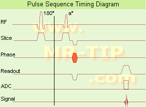
Ultrafast Gradient Echo Sequence Timing Diagram In simple ultrafast GRE imaging, TR and TE are so short, that tissues have a poor imaging signal and - more importantly - poor contrast except when contrast media enhanced (contrast enhanced angiography). Therefore, the magnetization is 'prepared' during the preparation module, most frequently by an initial 180° inversion pulse.
In the pulse sequence timing diagram, the basic ultrafast gradient echo sequence is illustrated. The 180° inversion pulse is executed one time (to the left of the vertical line), the right side represents the data collection period and is often repeated depending on the acquisition parameters.
See also Pulse Sequence Timing Diagram, there you will find a description of the components.
Ultrafast GRE sequences have a short TR,TE, a low flip angle and TR is so short that image acquisition lasts less than 1 second and typically less than 500 ms. Common TR: 3-5 msec, TE: 2 msec, and the flip angle is about 5°. Such sequences are often labeled with the prefix 'Turbo' like TurboFLASH, TurboFFE and TurboGRASS.
This allows one to center the subsequent ultrafast GRE data acquisition around the inversion time TI, where one of the tissues of interest has very little signal as its z-magnetization is passing through zero.
Unlike a standard inversion recovery (IR) sequence, all lines or a substantial segment of k-space image lines are acquired after a single inversion pulse, which can then together be considered as readout module. The readout module may use a variable flip angle approach, or the data acquisition may be divided into multiple segments (shots). The latter is useful particularly in cardiac imaging where acquiring all lines in a single segment may take too long relative to the cardiac cycle to provide adequate temporal resolution.
If multiple lines are acquired after a single pulse, the pulse sequence is a type of gradient echo echo planar imaging (EPI) pulse sequence.

See also Magnetization Prepared Rapid Gradient Echo (MPRAGE) and Turbo Field Echo (TFE).
spacer

• View the DATABASE results for 'Ultrafast Gradient Echo Sequence' (13).Open this link in a new window

Searchterm 'Second' was also found in the following services: 
spacer
Radiology  (26) Open this link in a new windowUltrasound  (49) Open this link in a new window
Ultrasmall Superparamagnetic Iron OxideInfoSheet: - Contrast Agents - 
Intro, Overview, 
Characteristics, 
Types of, 
etc.
 
(USPIO) The class of the ultrasmall superparamagnetic iron oxide includes several chemically and pharmacologically very distinct materials, which may or may not be interchangeable for a specific use. Some ultrasmall SPIO particles (median diameter less than 50nm) are used as MRI contrast agents (Sinerem®, Combidex®), e.g. to differentiate metastatic from inflammatory lymph nodes. USPIO shows also potential for providing important information about angiogenesis in cancer tumors and could possibly complement MRI helping physicians to identify dangerous arteriosclerosis plaques.
Because of the disadvantageous large T2*//T1 ratio, USPIO compounds are less suitable for arterial bolus contrast enhanced magnetic resonance angiography than gadolinium complexes. The tiny ultrasmall superparamagnetic iron oxides do not accumulate in the RES system as fast as larger particles, which results in a long plasma half-life. USPIO particles, with a small median diameter (less than 10 nm), will accumulate in lymph nodes after an intravenous injection by e.g. direct transcapillary passage through endothelial venules. Once within the nodal parenchyma, phagocytic cells of the mononuclear phagocyte system take up the particles.
As a second way, USPIOs are subsequently taken up from then interstitium by lymphatic vessels and transported to regional lymph nodes. A lymph node with normal phagocytic function takes up a considerable amount and shows a reduction of the signal intensity caused by T2 shortening effects and magnetic susceptibility. Caused by the small uptake of the USPIOs in metastatic lymph nodes, they appear with less signal reduction, and permit the differentiation of healthy lymph nodes from normal-sized, metastatic nodes.

See also Superparamagnetic Contrast Agents, Superparamagnetic Iron Oxide, Very Small Superparamagnetic Iron Oxide Particles, Blood Pool Agents, Intracellular Contrast Agents.
spacer

• View the DATABASE results for 'Ultrasmall Superparamagnetic Iron Oxide' (16).Open this link in a new window


• View the NEWS results for 'Ultrasmall Superparamagnetic Iron Oxide' (2).Open this link in a new window.
 
Further Reading:
  Basics:
Comparison of Two Superparamagnetic Viral-Sized Iron Oxide Particles Ferumoxides and Ferumoxtran-10 with a Gadolinium Chelate in Imaging Intracranial Tumors
2002   by www.ajnr.org    
  News & More:
Optimized Labelling of Human Monocytes with Iron Oxide MR Contrast Agents
Sunday, 30 November 2003   by rsna2003.rsna.org    
10 SUMMARY AND FUTURE PERSPECTIVES
   by dissertations.ub.rug.nl    
MRI Resources 
Equipment - MRI Training Courses - Contrast Agents - Stent - IR - Patient Information
 
previous      76 - 80 (of 83)     next
Result Pages : [1]  [2 3 4 5 6 7 8 9 10 11 12 13 14 15 16 17]
 Random Page
 
Share This Page
FacebookTwitterLinkedIn

MR-TIP    
Community   
User
Pass
Forgot your UserID/Password ?    


How AI will impact MRI :
only diagnostics 
saving time 
reducing cost 
makes planning obsolete 
reduce human knowledge 
not at all 

Look
      Ups


MR-TIP.com uses cookies! By browsing MR-TIP.com, you agree to our use of cookies.

Magnetic Resonance - Technology Information Portal
Member of SoftWays' Medical Imaging Group - MR-TIP • Radiology-TIP • Medical-Ultrasound-Imaging • 
Copyright © 2003 - 2024 SoftWays. All rights reserved. [ 25 April 2024]
Terms of Use | Privacy Policy | Advertising
 [last update: 2024-02-26 03:41:00]