Magnetic Resonance - Technology Information Portal Welcome to MRI Technology
Info
  Sheets

Out-
      side


 'Saturation' 
SEARCH FOR    
 
  2 3 5 A B C D E F G H I J K L M N O P Q R S T U V W X Y Z
Result : Searchterm 'Saturation' found in 12 terms [] and 41 definitions []
previous     11 - 15 (of 53)     next
Result Pages : [1 2 3]  [4 5 6 7 8 9 10 11]
Searchterm 'Saturation' was also found in the following services: 
spacer
News  (2)  Forum  (6)  
 
Spectral Presaturation Inversion Recovery
 
(SPIR) A specialized technique that selectively saturates fat protons prior to acquiring data as in standard sequences, so that they produce a negligible signal. The presaturation pulse is applied prior to each slice selection. This technique requires a very homogeneous magnetic field and very precise frequency calibration.
 
Images, Movies, Sliders:
 MRI Orbita T2 FatSat  Open this link in a new window
    
 
spacer
 
• Related Searches:
    • Inversion Recovery Sequence
    • Prepulse
    • Abdominal Imaging
    • Fat Suppression
    • Image Contrast Characteristics
 
Further Reading:
  Basics:
Sequence for Philips(.pdf)
   by www.droid.cuhk.edu.hk    
Optimizing SPIR and SPAIR fat suppression
Tuesday, 30 November 2004   by clinical.netforum.healthcare.philips.com    
Techniques of Fat Suppression(.pdf)
   by cds.ismrm.org    
Searchterm 'Saturation' was also found in the following service: 
spacer
Ultrasound  (5) Open this link in a new window
Paramagnetic Chemical Exchange Saturation TransferInfoSheet: - Contrast Agents - 
Intro, Overview, 
Characteristics, 
Types of, 
etc.
 
(PARACEST) The alteration of the proton density or total water signal changes contrast and can be detected by the MRI scanner. Paramagnetic chemical exchange saturation transfer contrast agents are based upon the magnetization transfer mechanism.
Lanthanide ion complexes formed with tetra-amide based ligands display unusually slow water exchange kinetics and this feature may be used to alter image contrast by applying a selective presaturation pulse in an imaging sequence. This results in chemical exchange saturation transfer (CEST) from the lanthanide-bound water to bulk water thereby altering image contrast.
Chemical Exchange Saturation Transfer (CEST) agents are a class of contrast agents that could potentially revolutionize the MRI field because of their improved sensitivity and can have a great impact on functional magnetic resonance imaging.
spacer
 
Further Reading:
  Basics:
Multimodal Nanoparticles for Quantitative Imaging(.pdf)
Tuesday, 13 December 2011   by alexandria.tue.nl    
  News & More:
New Brain Imaging Technique Identifies Previously Undetected Epileptic Seizure Sites
Friday, 13 November 2015   by www.newswise.com    
Non-invasive Imaging Method For Diagnosing Osteoarthritis Developed
Friday, 15 February 2008   by www.sciencedaily.com    
MRI Resources 
Functional MRI - Guidance - Software - Bioinformatics - Blood Flow Imaging - Calculation
 
Gradient Echo SequenceForum -
related threadsInfoSheet: - Sequences - 
Intro, 
Overview, 
Types of, 
etc.
 
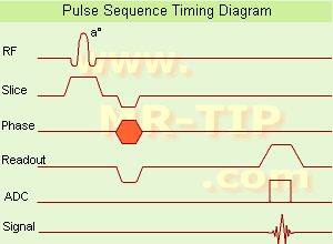
Gradient Echo Sequence Timing Diagram (GRE - sequence) A gradient echo is generated by using a pair of bipolar gradient pulses. In the pulse sequence timing diagram, the basic gradient echo sequence is illustrated. There is no refocusing 180° pulse and the data are sampled during a gradient echo, which is achieved by dephasing the spins with a negatively pulsed gradient before they are rephased by an opposite gradient with opposite polarity to generate the echo.
See also the Pulse Sequence Timing Diagram. There you will find a description of the components.
The excitation pulse is termed the alpha pulse α. It tilts the magnetization by a flip angle α, which is typically between 0° and 90°. With a small flip angle there is a reduction in the value of transverse magnetization that will affect subsequent RF pulses. The flip angle can also be slowly increased during data acquisition (variable flip angle: tilt optimized nonsaturation excitation). The data are not acquired in a steady state, where z-magnetization recovery and destruction by ad-pulses are balanced. However, the z-magnetization is used up by tilting a little more of the remaining z-magnetization into the xy-plane for each acquired imaging line.
Gradient echo imaging is typically accomplished by examining the FID, whereas the read gradient is turned on for localization of the signal in the readout direction. T2* is the characteristic decay time constant associated with the FID. The contrast and signal generated by a gradient echo depend on the size of the longitudinal magnetization and the flip angle. When α = 90° the sequence is identical to the so-called partial saturation or saturation recovery pulse sequence. In standard GRE imaging, this basic pulse sequence is repeated as many times as image lines have to be acquired. Additional gradients or radio frequency pulses are introduced with the aim to spoil to refocus the xy-magnetization at the moment when the spin system is subject to the next α pulse.
As a result of the short repetition time, the z-magnetization cannot fully recover and after a few initial α pulses there is an equilibrium established between z-magnetization recovery and z-magnetization reduction due to the α pulses.
Gradient echoes have a lower SAR, are more sensitive to field inhomogeneities and have a reduced crosstalk, so that a small or no slice gap can be used. In or out of phase imaging depending on the selected TE (and field strength of the magnet) is possible. As the flip angle is decreased, T1 weighting can be maintained by reducing the TR. T2* weighting can be minimized by keeping the TE as short as possible, but pure T2 weighting is not possible. By using a reduced flip angle, some of the magnetization value remains longitudinal (less time needed to achieve full recovery) and for a certain T1 and TR, there exist one flip angle that will give the most signal, known as the "Ernst angle".
Contrast values:
PD weighted: Small flip angle (no T1), long TR (no T1) and short TE (no T2*)
T1 weighted: Large flip angle (70°), short TR (less than 50ms) and short TE
T2* weighted: Small flip angle, some longer TR (100 ms) and long TE (20 ms)

Classification of GRE sequences can be made into four categories:
See also Gradient Recalled Echo Sequence, Spoiled Gradient Echo Sequence, Refocused Gradient Echo Sequence, Ultrafast Gradient Echo Sequence.
 
Images, Movies, Sliders:
 MRI Liver In Phase  Open this link in a new window
    
 MRI Liver Out Of Phase  Open this link in a new window
    
 MVP Parasternal  Open this link in a new window
 Breast MRI Images T1 Pre - Post Contrast  Open this link in a new window
 Circle of Willis, Time of Flight, MIP  Open this link in a new window
    
SlidersSliders Overview

 
spacer

• View the DATABASE results for 'Gradient Echo Sequence' (70).Open this link in a new window

 
Further Reading:
  Basics:
Enhanced Fast GRadient Echo 3-Dimensional (efgre3D) or THRIVE
   by www.mri.tju.edu    
  News & More:
MRI evaluation of fatty liver in day to day practice: Quantitative and qualitative methods
Wednesday, 3 September 2014   by www.sciencedirect.com    
T1rho-prepared balanced gradient echo for rapid 3D T1rho MRI
Monday, 1 September 2008   by www.ncbi.nlm.nih.gov    
Searchterm 'Saturation' was also found in the following services: 
spacer
News  (2)  Forum  (6)  
 
Black Blood MRAForum -
related threadsInfoSheet: - Sequences - 
Intro, 
Overview, 
Types of, 
etc.MRI Resource Directory:
 - Cardiovascular Imaging -
 
With this magnetic resonance angiography technique flowing blood appears dark.
MR black blood techniques have been developed for cardiovascular imaging to improve segmentation of myocardium from the blood pool. Black blood MRA techniques decrease the signal from blood with reference to the myocardium and make it easier to perform cardiac chamber segmentation.
ECG gated spin echo sequences with presaturation pulses for magnetization preparation will show strong intravascular signal loss due to flow effects when appropriate imaging conditions including spatial presaturation are used. The sequence use the flow void effect as blood passes rapidly through the selected slice.
For dark blood preparation, a pair of nonselective and selective 180° inversion pulses are used, followed by a long inversion time to null signal from inflowing blood. A second selective inversion pulse can also be applied with short inversion time to null the fat signal. These in cardiac imaging used black blood techniques are referred to as double inversion recovery T1 measurement turbo spin echo or fast spin echo, and double-inversion recovery STIR.
 
Images, Movies, Sliders:
 Normal Dual Inversion Fast Spin-echo  Open this link in a new window
      

Courtesy of  Robert R. Edelman

 
spacer

• View the DATABASE results for 'Black Blood MRA' (6).Open this link in a new window

Searchterm 'Saturation' was also found in the following service: 
spacer
Ultrasound  (5) Open this link in a new window
Fat SuppressionForum -
related threads
 
Fat suppression is the process of utilizing specific MRI parameters to remove the deleterious effects of fat from the resulting images , e.g. with STIR, FAT SAT sequences, water selective (PROSET WATS - water only selection, also FATS - fat only selection possible) excitation techniques, or pulse sequences based on the Dixon method.
Spin magnetization can be modulated by using special RF pulses. CHESS or its variations like SPIR, SPAIR (Spectral Selection Attenuated Inversion Recovery) and FAT SAT use frequency selective excitation pulses, which produce fat saturation.
Fat suppression techniques are nearly used in all body parts and belong to every standard MRI protocol of joints like knee, shoulder, hips, etc.
mri safety guidance
Image Guidance
Imaging of, e.g. the foot can induce bad fat suppression with SPIR/FAT SAT due to the asymmetric volume of this body part. The volume of the foot alters the magnetic field to a different degree than the smaller volume of the lower leg affecting the protons there. There is only a small band of tissue where the fat protons are precessing at the frequency expected, resulting in frequency selective fat saturation working only in that area. This can be corrected by volume shimming or creating a more symmetrical volume being imaged with water bags.
Even with their longer scan time and motion sensitivity, STIR (short T1/tau inversion recovery) sequences are often the better choice to suppress fat. STIR images are also preferred because of the decreased sensitivity to field inhomogeneities, permitting larger fields of views when compared to fat suppressed images and the ability to image away from the isocenter.
See also Knee MRI.
Sequences based on Dixon turbo spin echo (fast spin echo) can deliver a significant better fat suppression than conventional TSE/FSE imaging.
 
Images, Movies, Sliders:
 Shoulder Axial T2 FatSat FRFSE  Open this link in a new window
    

Courtesy of  Robert R. Edelman
 MRI Orbita T2 FatSat  Open this link in a new window
    
 Knee MRI Sagittal STIR 001  Open this link in a new window
 MRI - Anatomic Imaging of the Ankle 3  Open this link in a new window
    
SlidersSliders Overview

 
spacer

• View the DATABASE results for 'Fat Suppression' (28).Open this link in a new window

 
Further Reading:
  Basics:
Techniques of Fat Suppression(.pdf)
   by cds.ismrm.org    
  News & More:
Enhanced Fast GRadient Echo 3-Dimensional (efgre3D) or THRIVE
   by www.mri.tju.edu    
Ultrashort echo time (UTE) MRI of the spine in thalassaemia
February 2004   by bjr.birjournals.org    
MRI Resources 
Intraoperative MRI - Fluorescence - Safety Products - Quality Advice - Patient Information - Contrast Agents
 
previous      11 - 15 (of 53)     next
Result Pages : [1 2 3]  [4 5 6 7 8 9 10 11]
 Random Page
 
Share This Page
FacebookTwitterLinkedIn

MR-TIP    
Community   
User
Pass
Forgot your UserID/Password ?    


New acceleration techniques will :
reduce scan times 
cause artifacts 
increase expenses 
be useful if you have a lot of experience 
doesn't do much 
never heard of 

Look
      Ups


MR-TIP.com uses cookies! By browsing MR-TIP.com, you agree to our use of cookies.

Magnetic Resonance - Technology Information Portal
Member of SoftWays' Medical Imaging Group - MR-TIP • Radiology-TIP • Medical-Ultrasound-Imaging • 
Copyright © 2003 - 2024 SoftWays. All rights reserved. [ 19 April 2024]
Terms of Use | Privacy Policy | Advertising
 [last update: 2024-02-26 03:41:00]