Magnetic Resonance - Technology Information Portal Welcome to MRI Technology
Info
  Sheets

Out-
      side


 'Saturation%20Recovery' 
SEARCH FOR    
 
  2 3 5 A B C D E F G H I J K L M N O P Q R S T U V W X Y Z
Result : Searchterm 'Saturation Recovery' found in 1 term [] and 4 definitions [], (+ 8 Boolean[] results
1 - 5 (of 13)     next
Result Pages : [1]  [2 3]
MRI Resources 
Databases - Contrast Enhanced MRI - Implant and Prosthesis pool - Open Directory Project - Spine MRI - Shielding
 
Saturation Recovery
 
(SR) Particular type of partial saturation pulse sequence in which the preceding pulses leave the spins in a state of saturation, so that recovery at the time of the next pulse has taken place from an initial condition of no magnetization. A rare used MRI pulse sequence that generates a predominantly proton density dependent signal, basically employing a 90° RF excitation pulse, with a very long repetition time. With this technique T1 times can be measured faster than with inversion recovery pulse sequences.
This saturation recovery sequence consists of multiple 90° radio frequency (RF) pulses with a short repetition time. A spoiler gradient pulse dephases the longitudinal magnetization that remains after the first 90° radio frequency pulse. A repetition time interval after the application of this spoiling gradient turns an additional 90° pulse the new developed longitudinal magnetization into the transverse plane, followed by recording a gradient echo.
spacer
 
• Share the entry 'Saturation Recovery':  Facebook  Twitter  LinkedIn  
 
• Related Searches:
    • Saturation
    • Progressive Saturation
    • Fat Saturation
    • Suppression
    • Spectral Presaturation Inversion Recovery
 
Further Reading:
  Basics:
Contrast mechanisms in magnetic resonance imaging
2004   by www.iop.org    
MRI Resources 
Stimulator pool - Patient Information - Sequences - MRI Accidents - Functional MRI - Spine MRI
 
Partial SaturationInfoSheet: - Sequences - 
Intro, 
Overview, 
Types of, 
etc.
 
(PS) Excitation technique applying repeated RF pulses in times comparable to or shorter than T1. Incomplete T1 relaxation leads to reduction of the signal amplitude; there is the possibility of generating images with increased contrast between regions with different relaxation times.
Although partial saturation is also commonly referred to as saturation recovery, that term should properly be reserved for the particular case of partial saturation in which recovery after each excitation effectively takes place from true saturation. A GRE sequence where α = 90° is identical to the partial saturation or saturation recovery pulse sequence.
It does not directly produce images of T1. However, since the measured signal will depend on T1, the method generates contrast between regions with different relaxation times. If T2 and/or T2 effects are minimized through the use of a short echo time TE, the result is a T1 weighted image. It is not a T1 image due to the possible presence of spin density and T2 effects as well as the nonlinear dependence on T1.
The change in signal from a region resulting from a change in the interpulse time, TR, can be used to calculate T1 for the region.
spacer

• View the DATABASE results for 'Partial Saturation' (5).Open this link in a new window

MRI Resources 
Spine MRI - Manufacturers - General - Software - - Intraoperative MRI
 
Gradient Echo SequenceForum -
related threadsInfoSheet: - Sequences - 
Intro, 
Overview, 
Types of, 
etc.
 
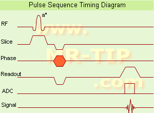
Gradient Echo Sequence Timing Diagram (GRE - sequence) A gradient echo is generated by using a pair of bipolar gradient pulses. In the pulse sequence timing diagram, the basic gradient echo sequence is illustrated. There is no refocusing 180° pulse and the data are sampled during a gradient echo, which is achieved by dephasing the spins with a negatively pulsed gradient before they are rephased by an opposite gradient with opposite polarity to generate the echo.
See also the Pulse Sequence Timing Diagram. There you will find a description of the components.
The excitation pulse is termed the alpha pulse α. It tilts the magnetization by a flip angle α, which is typically between 0° and 90°. With a small flip angle there is a reduction in the value of transverse magnetization that will affect subsequent RF pulses. The flip angle can also be slowly increased during data acquisition (variable flip angle: tilt optimized nonsaturation excitation). The data are not acquired in a steady state, where z-magnetization recovery and destruction by ad-pulses are balanced. However, the z-magnetization is used up by tilting a little more of the remaining z-magnetization into the xy-plane for each acquired imaging line.
Gradient echo imaging is typically accomplished by examining the FID, whereas the read gradient is turned on for localization of the signal in the readout direction. T2* is the characteristic decay time constant associated with the FID. The contrast and signal generated by a gradient echo depend on the size of the longitudinal magnetization and the flip angle. When α = 90° the sequence is identical to the so-called partial saturation or saturation recovery pulse sequence. In standard GRE imaging, this basic pulse sequence is repeated as many times as image lines have to be acquired. Additional gradients or radio frequency pulses are introduced with the aim to spoil to refocus the xy-magnetization at the moment when the spin system is subject to the next α pulse.
As a result of the short repetition time, the z-magnetization cannot fully recover and after a few initial α pulses there is an equilibrium established between z-magnetization recovery and z-magnetization reduction due to the α pulses.
Gradient echoes have a lower SAR, are more sensitive to field inhomogeneities and have a reduced crosstalk, so that a small or no slice gap can be used. In or out of phase imaging depending on the selected TE (and field strength of the magnet) is possible. As the flip angle is decreased, T1 weighting can be maintained by reducing the TR. T2* weighting can be minimized by keeping the TE as short as possible, but pure T2 weighting is not possible. By using a reduced flip angle, some of the magnetization value remains longitudinal (less time needed to achieve full recovery) and for a certain T1 and TR, there exist one flip angle that will give the most signal, known as the "Ernst angle".
Contrast values:
PD weighted: Small flip angle (no T1), long TR (no T1) and short TE (no T2*)
T1 weighted: Large flip angle (70°), short TR (less than 50ms) and short TE
T2* weighted: Small flip angle, some longer TR (100 ms) and long TE (20 ms)

Classification of GRE sequences can be made into four categories:
See also Gradient Recalled Echo Sequence, Spoiled Gradient Echo Sequence, Refocused Gradient Echo Sequence, Ultrafast Gradient Echo Sequence.
 
Images, Movies, Sliders:
 MRI Liver In Phase  Open this link in a new window
    
 MRI Liver Out Of Phase  Open this link in a new window
    
 MVP Parasternal  Open this link in a new window
 Breast MRI Images T1 Pre - Post Contrast  Open this link in a new window
 Circle of Willis, Time of Flight, MIP  Open this link in a new window
    
SlidersSliders Overview

 
spacer

• View the DATABASE results for 'Gradient Echo Sequence' (70).Open this link in a new window

 
Further Reading:
  Basics:
Enhanced Fast GRadient Echo 3-Dimensional (efgre3D) or THRIVE
   by www.mri.tju.edu    
  News & More:
MRI evaluation of fatty liver in day to day practice: Quantitative and qualitative methods
Wednesday, 3 September 2014   by www.sciencedirect.com    
T1rho-prepared balanced gradient echo for rapid 3D T1rho MRI
Monday, 1 September 2008   by www.ncbi.nlm.nih.gov    
MRI Resources 
Spine MRI - Resources - Hospitals - Developers - Mobile MRI - DICOM
 
Progressive Saturation
 
spacer
MRI Resources 
Safety Products - MRI Physics - Knee MRI - MRI Training Courses - Developers - Guidance
 
Saturation
 
In MR, saturation is a nonequilibrium state with no net magnetization. The same amount of nuclear spins is aligned against and with the magnetic field. Saturation methods like FatSat, SPIR etc., work with a frequency selective saturation pulse for a specific chemical shift applied before the actual sequence starts. This saturation pulse adjusts the magnetization from tissue components to zero. The hydrogen nuclei of fat and water resonate at different frequencies, which makes it possible to excite just the fat with repeatedly applying RF pulses at the Larmor frequency with interpulse times compared to T1. The resulting signal is then destroyed with a gradient pulse (Spoiler Gradient Pulse). Fat is the chemical compound to be saturated at a fat saturation sequence. When the actual sequence follows, (e.g., a spin echo sequence) the unwanted suppressed component will not resonate.

See also Saturation Recovery.
spacer

• View the DATABASE results for 'Saturation' (54).Open this link in a new window

MRI Resources 
Equipment - Health - General - Calculation - Spectroscopy - Coils
 
     1 - 5 (of 13)     next
Result Pages : [1]  [2 3]
 Random Page
 
Share This Page
FacebookTwitterLinkedIn

MR-TIP    
Community   
User
Pass
Forgot your UserID/Password ?    


New acceleration techniques will :
reduce scan times 
cause artifacts 
increase expenses 
be useful if you have a lot of experience 
doesn't do much 
never heard of 

Look
      Ups


MR-TIP.com uses cookies! By browsing MR-TIP.com, you agree to our use of cookies.

Magnetic Resonance - Technology Information Portal
Member of SoftWays' Medical Imaging Group - MR-TIP • Radiology-TIP • Medical-Ultrasound-Imaging • 
Copyright © 2003 - 2024 SoftWays. All rights reserved. [ 20 April 2024]
Terms of Use | Privacy Policy | Advertising
 [last update: 2024-02-26 03:41:00]