Magnetic Resonance - Technology Information Portal Welcome to MRI Technology
Info
  Sheets

Out-
      side


 'Repetition Time' 
SEARCH FOR    
 
  2 3 5 A B C D E F G H I J K L M N O P Q R S T U V W X Y Z
Result : Searchterm 'Repetition Time' found in 1 term [] and 32 definitions []
previous     11 - 15 (of 33)     next
Result Pages : [1]  [2 3 4 5 6 7]
Searchterm 'Repetition Time' was also found in the following services: 
spacer
News  (1)  Forum  (1)  
 
Echo Planar ImagingInfoSheet: - Sequences - 
Intro, 
Overview, 
Types of, 
etc.MRI Resource Directory:
 - Sequences -
 
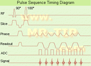
Echo Planar Imaging Timing Diagram (EPI) Echo planar imaging is one of the early magnetic resonance imaging sequences (also known as Intascan), used in applications like diffusion, perfusion, and functional magnetic resonance imaging. Other sequences acquire one k-space line at each phase encoding step. When the echo planar imaging acquisition strategy is used, the complete image is formed from a single data sample (all k-space lines are measured in one repetition time) of a gradient echo or spin echo sequence (see single shot technique) with an acquisition time of about 20 to 100 ms. The pulse sequence timing diagram illustrates an echo planar imaging sequence from spin echo type with eight echo train pulses. (See also Pulse Sequence Timing Diagram, for a description of the components.)
In case of a gradient echo based EPI sequence the initial part is very similar to a standard gradient echo sequence. By periodically fast reversing the readout or frequency encoding gradient, a train of echoes is generated.
EPI requires higher performance from the MRI scanner like much larger gradient amplitudes. The scan time is dependent on the spatial resolution required, the strength of the applied gradient fields and the time the machine needs to ramp the gradients.
In EPI, there is water fat shift in the phase encoding direction due to phase accumulations. To minimize water fat shift (WFS) in the phase direction fat suppression and a wide bandwidth (BW) are selected. On a typical EPI sequence, there is virtually no time at all for the flat top of the gradient waveform. The problem is solved by "ramp sampling" through most of the rise and fall time to improve image resolution.
The benefits of the fast imaging time are not without cost. EPI is relatively demanding on the scanner hardware, in particular on gradient strengths, gradient switching times, and receiver bandwidth. In addition, EPI is extremely sensitive to image artifacts and distortions.
spacer
 
• Related Searches:
    • Perfusion Imaging
    • K-Space Trajectory
    • Blipped Phase Encoding
    • Motion Probing Gradient
    • Functional Magnetic Resonance Imaging
 
Further Reading:
  Basics:
New Imaging Method Makes Brain Scans 7 Times Faster
Sunday, 9 January 2011   by www.dailytech.com    
MRI Resources 
MR Guided Interventions - Journals - Brain MRI - Stent - Movies - Education pool
 
Echo TimeForum -
related threads
 
(TE) The echo time represents the time in milliseconds between the application of the 90° pulse and the peak of the echo signal in spin echo and inversion recovery pulse sequences.

See also Repetition Time, Spin Echo Sequence and Inversion Recovery Sequence.
spacer

• View the DATABASE results for 'Echo Time' (36).Open this link in a new window

 
Further Reading:
  Basics:
Magnetic resonance imaging
   by www.scholarpedia.org    
  News & More:
Kidney stone imaging with 3D ultra-short echo time (UTE) magnetic resonance imaging. A phantom study - Abstract
Wednesday, 11 March 2015   by www.urotoday.com    
Ultrashort echo time (UTE) MRI of the spine in thalassaemia
February 2004   by bjr.birjournals.org    
MRI Resources 
Nerve Stimulator - Lung Imaging - Movies - MRI Training Courses - Mass Spectrometry -
 
Fast Relaxation Fast Spin EchoInfoSheet: - Sequences - 
Intro, 
Overview, 
Types of, 
etc.
 
(FRFSE, FR-FSE) The fast relaxation fast spin echo sequence provides high signal intensity of fluids even with short repetition times, and can be used with parallel imaging techniques for short breath hold imaging or respiratory gating for free-breathing, high isotropic resolution MR imaging. After signal decay at the end of the echo train, a negative 90° pulse align spins with long T2 from the transverse plane to the longitudinal plane, leading to a much faster recovery of tissues with long T2 time to the equilibrium and thus better contrast between tissues with long and short T2.
Fast relaxation FSE has advantages also for volumetric imaging as the TR can be substantially reduced and thus the scan time. The sequence can be post processed with maximum intensity projection, surface or volume rendering algorithms to visualize anatomical details in brain or spine MRI. Cerebro spinal fluid pulsation artifacts, often problematic in the cervical or thoracic spine may be reduced by radial sampling, in particular when combined with acquisitions of the PROPELLER type.

See also Fast spin echo, Driven Equilibrium.
 
Images, Movies, Sliders:
 Shoulder Sagittal T2 FatSat FRFSE  Open this link in a new window
    

Courtesy of  Robert R. Edelman
 Shoulder Axial T2 FatSat FRFSE  Open this link in a new window
 Shoulder Coronal T2 FatSat FRFSE  Open this link in a new window
    

Courtesy of  Robert R. Edelman
 
spacer
Searchterm 'Repetition Time' was also found in the following services: 
spacer
News  (1)  Forum  (1)  
 
Flow ArtifactInfoSheet: - Artifacts - 
Case Studies, 
Reduction Index, 
etc.MRI Resource Directory:
 - Artifacts -
 
Quick Overview
Please note that there are different common names for this artifact.
Artifact Information
DESCRIPTION
Vascular ghosts (ghosting artifact), anomalous intensities in images
REASON
Movement of body fluids
HELP
Flow compensation, presaturation, triggering
Flow effects in MRI produce a range of artifacts, e.g. intravascular signal void by time of flight effects; turbulent dephasing and first echo dephasing, caused by flowing blood.
Through movement of the hydrogen nuclei (e.g. blood flow), there is a location change between the time these nuclei experience a radio frequency pulse and the time the emitted signal is received (because the repetition time is asynchronous with the pulsatile flow).
The blood flow occasionally produces intravascular high signal intensities due to flow related enhancement, even echo rephasing and diastolic pseudogating. The pulsatile laminar flow within vessels often produces a complex multilayered band that usually propagates outside the head in the phase encoded direction. Blood flow artifacts should be considered as a special subgroup of motion artifacts.
mri safety guidance
Image Guidance
Artifacts can be reduced by reduction of phase shifts with flow compensation (gradient moment nulling), suppression of the blood signal with saturation pulses parallel to the slices, synchronization of the imaging sequence with the heart cycle (cardiac triggering) or can be flipped 90° by swapping the phase//frequency encoding directions.

See also Flow Related Enhancement and Flow Effects.
 
Images, Movies, Sliders:
 Knee MRI Sagittal T1 003  Open this link in a new window
 
spacer

• View the DATABASE results for 'Flow Artifact' (6).Open this link in a new window

 
Further Reading:
  News & More:
MRI measure of blood flow over atherosclerotic plaque may detect dangerous plaque
Friday, 5 April 2013   by www.sciencecodex.com    
Advanced Visualization Techniques Could Change the Paradigm for Diagnosis and Treatment of Heart Disease
Thursday, 31 May 2012   by www.sciencedaily.com    
MRI Resources 
MRI Technician and Technologist Schools - Databases - Artifacts - Spectroscopy pool - - Contrast Agents
 
Flow Related Enhancement
 
(FRE) Flow related enhancement could be seen most for blood flow, but also for other liquids with some MR imaging techniques, as an increase in intensity due to the washout of saturated spins. FRE provides positive contrast ("bright blood") of vascular details in time of flight MRA as well as the physiologic characterization of blood flow.
If stationary spins within the scanned region experience only an incomplete T1 relaxation between the repeated radio frequency (RF) excitations, this results in fewer signal of the stationary tissue (compared to inflowing blood with completely relaxed spins). The degree of the flow related enhancement is proportional to the blood flow velocity and the used repetition time. The use of flow compensation (gradient moment nulling) improves the FRE especially in gradient echo sequences.
 
Images, Movies, Sliders:
 TOF-MRA Circle of Willis Inverted MIP  Open this link in a new window
    

 Circle of Willis, Time of Flight, MIP  Open this link in a new window
    
SlidersSliders Overview

 
spacer

• View the DATABASE results for 'Flow Related Enhancement' (10).Open this link in a new window

 
Further Reading:
  Basics:
Conventional MRI and MR Angiography of Stroke
2012   by www.mc.vanderbilt.edu    
MRI Resources 
Pathology - Chemistry - MR Myelography - Absorption and Emission - Spine MRI - Manufacturers
 
previous      11 - 15 (of 33)     next
Result Pages : [1]  [2 3 4 5 6 7]
 Random Page
 
Share This Page
FacebookTwitterLinkedIn

MR-TIP    
Community   
User
Pass
Forgot your UserID/Password ?    


MRI is trending to low field magnets :
reduced costs will lead to this change 
AI will close the gap to high field 
only in remote areas 
is only temporary 
never 

Look
      Ups


MR-TIP.com uses cookies! By browsing MR-TIP.com, you agree to our use of cookies.

Magnetic Resonance - Technology Information Portal
Member of SoftWays' Medical Imaging Group - MR-TIP • Radiology-TIP • Medical-Ultrasound-Imaging • 
Copyright © 2003 - 2024 SoftWays. All rights reserved. [ 23 April 2024]
Terms of Use | Privacy Policy | Advertising
 [last update: 2024-02-26 03:41:00]