 | Info
Sheets |
| | | | | | | | | | | | | | | | | | | | | | | | |
 | Out-
side |
| | | | |
|
| | | | |
Result : Searchterm 'Real' found in 4 terms [ ] and 43 definitions [ ] and 43 definitions [ ] ]
| | previous 41 - 45 (of 47) nextResult Pages : [1] [2 3 4 5 6 7 8 9 10] |  | |  | Searchterm 'Real' was also found in the following services: | | | | |
| |  |
| |
|

(Signa VH/i 3.0T)
With GE Healthcare
leading-edge technology in ultra-high-field imaging. The 3 T VH/i provides a platform for advanced applications in radiology, cardiology, psychology and psychiatry. Real-time image processing lets you acquire multislice whole brain images and map brain functions for research or surgical planning. And the 3 T Signa VH/i is flexible enough to provide clinicians with high performance they require. It can provide not only outstanding features in brain scanning and neuro-system research, but also a wide range of use in scanning breasts, extremities, the spine and the cardiovascular systems.
Device Information and Specification CLINICAL APPLICATION Whole body
T/R quadrature head, T/R quadrature body, T/R phased array extremity (opt) SE, IR, 2D/3D GRE, FGRE, RF-spoiled GRE, FSE, Angiography: 2D/3D TOF, 2D/3D phase contrast vascular IMAGING MODES Single, multislice, volume study, fast scan, multi slab, cine, localizer 100 Images/sec with Reflex100 MULTISLICE 100 Images/sec with Reflex100 2D 0.5-100mm in 0.1mm incremental 128x512 steps 32 phase encode H*W*D 260cm x 238cm x 265cm POWER REQUIREMENTS 480 or 380/415, 3 phase ||
COOLING SYSTEM TYPE Closed-loop water-cooled grad. Less than 0.14 L/hr liquid He | | | | | |
| | | Searchterm 'Real' was also found in the following services: | | | | |
| | |
| |
|
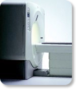
From GE Healthcare;
GE's Signa Contour/i system uses the innovations like K4 technology and real-time interactive imaging.
This compact magnet with wide-flare gantry obtains high patient comfort with low costs.
Device Information and Specification CLINICAL APPLICATION Whole body Head and body coil standard; all other coils optional; open architecture makes system compatible with a wide selection of coils Standard: SE, IR, 2D/3D GRE and SPGR, Angiography;; 2D/3D TOF, 2D/3D Phase Contrast;; 2D/3D FSE, 2D/3D FGRE and FSPGR, SSFP, FLAIR, optional: EPI, 2D/3D Fiesta, FGRET, Spiral2D 0.8 mm to 20 mm; 3D 0.1 mm to 5 mm 128x512 steps 32 phase encode POWER REQUIREMENTS 480 or 380/415 V STRENGTH SmartSpeed 23 mT/m, HiSpeed Plus 33 mT/m | | | | | |
| | | | | |
| |
|

From GE Healthcare;
The Signa HDx MRI system is GE's leading edge whole body magnetic resonance scanner designed to support high resolution, high signal to noise ratio, and short scan times.
Signa HDx 3.0T offers new technologies like ultra-fast image reconstruction through the new XVRE recon engine, advancements in parallel imaging algorithms and the broadest range of premium applications. The HD applications, PROPELLER (high-quality brain imaging extremely resistant to motion artifacts), TRICKS (contrast-enhanced angiographic vascular lower leg imaging), VIBRANT (for breast MRI), LAVA (high resolution liver imaging with shorter breath holds and better organ coverage) and MR Echo (high-definition cardiac images in real time) offer unique capabilities.
Device Information and Specification CLINICAL APPLICATION Whole body
CONFIGURATION Compact short bore SE, IR, 2D/3D GRE, RF-spoiled GRE, 2DFGRE, 2DFSPGR, 3DFGRE, 3DFSPGR, 3DTOFGRE, 3DFSPGR, 2DFSE, 2DFSE-XL, 2DFSE-IR, T1-FLAIR, SSFSE, EPI, DW-EPI, BRAVO, Angiography: 2D/3D TOF, 2D/3D phase contrast vascular IMAGING MODES Single, multislice, volume study, fast scan, multi slab, cine, localizer H*W*D 240 x 2216,6 x 201,6 cm POWER REQUIREMENTS 480 or 380/415, 3 phase ||
COOLING SYSTEM TYPE Closed-loop water-cooled grad. | | | | | |
| | | Searchterm 'Real' was also found in the following services: | | | | |
| | | | | Searchterm 'Real' was also found in the following services: | | | | |
| | |
| |
|
A spectral line is a particular distinct frequency or narrow band of a frequency set. The resonance of this frequency occurs corresponding to a particular chemical shift. Theoretically, the frequency of a pure sine wave displays sharp spectral lines at the point of Larmor frequency. In reality, the spectral lines spread into a blurred peak, caused by field inhomogeneities and spin-spin effect. | | | |
• View the DATABASE results for 'Spectral Line' (21).
| | | | |
| | | | |
| | |
| | | |
|
| |
 | Look
Ups |
| |


