Magnetic Resonance - Technology Information Portal Welcome to MRI Technology
Info
  Sheets

Out-
      side


 'Pixel' 
SEARCH FOR    
 
  2 3 5 A B C D E F G H I J K L M N O P Q R S T U V W X Y Z
Result : Searchterm 'Pixel' found in 1 term [] and 66 definitions []
previous     26 - 30 (of 67)     next
Result Pages : [1]  [2 3 4 5 6 7 8 9 10 11 12 13 14]
Searchterm 'Pixel' was also found in the following services: 
spacer
Forum  (5)  
 
Gradient Motion Rephasing
 
(GMR) The application of strategic gradient pulses can compensate the objectionable spin phase effects of flow motion. That means the reducing of flow effects, e.g. gradient moment nulling of the first order of flow. The simplest velocity-compensated pulse sequence is the symmetrical second echo of a spin echo pulse sequence.
Gradient field changes can be configured in such a way that during an echo the magnetization signal vectors for all pixels have zero phase angle independent of velocities, accelerations etc. of the measured tissue. E.g. the adjustment to zero at the time TE of the net moments of the amplitude of the waveform of the magnetic field gradients with time. The zeroth moment is the area under the curve, the first moment is the 'center of gravity' etc. The aim is to minimize the phase shifts acquired by the transverse magnetization of excited nuclei moving along the gradients (including the effect of refocusing RF pulses), particularly for the reduction of image artifacts due to motion.
Also called Flow Compensation (FC), Motion Artifact Suppression Technique (MAST), Flow motion compression (STILL), Gradient Rephasing (GR), Shimadzu Motion Artifact Reduction Technique (SMART).
spacer
 
Further Reading:
  Basics:
Motion Compensation in MR Imaging
   by ccn.ucla.edu    
Searchterm 'Pixel' was also found in the following services: 
spacer
Radiology  (15) Open this link in a new windowUltrasound  (8) Open this link in a new window
Image Resolution
 
The image resolution is the level of detail of an image and a measurement of image quality. Higher resolution means more image detail, for example when two structures 1 mm apart are distinguishable in an image, this picture has a higher resolution than an image where they are not to distinguish.
More data points in an MR image (with same FOV) will decrease the pixel size, but not accurately improve the resolution because the different MRI sequences influence the contrast and the discernment of different tissues. With high contrast and optimal signal to noise ratio, the image resolution is depend on FOV and number of data points of a picture, but T2* effects have an additional influence.
spacer

• View the DATABASE results for 'Image Resolution' (9).Open this link in a new window


• View the NEWS results for 'Image Resolution' (1).Open this link in a new window.
 
Further Reading:
  Basics:
The Basics of MRI
   by www.cis.rit.edu    
  News & More:
Optimizing Musculoskeletal MR
   by rad.usuhs.mil    
MRI Resources 
Supplies - Spectroscopy pool - RIS - Cochlear Implant - Devices - DICOM
 
Infinion 1.5TPanorama 0.2InfoSheet: - Devices -
Intro, 
Types of Magnets, 
Overview, 
etc.MRI Resource Directory:
 - Devices -
 
From Philips Medical Systems; www.medical.philips.com/main/products/mri/products/infinion1.5t/features/ 
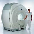
Philips Infinion 1.5 T is designed to maximize the efficiency and quality of patient care. Developed with the patient in mind, the Infinion is the shortest and most open 1.5T scanner available. The unique 'ultra short' 1.4 m magnet assures patient comfort and acceptance without compromising image quality and clinical performance.
Device Information and Specification
CLINICAL APPLICATION
Whole body
CONFIGURATION
Ultra short bore
Head, head / neck, integrated C-spine, L/T spine array, small large GP coils, body flex array, torso pelvis array, breast array, endocavitary, shoulder array, lower extremity, hand / wrist, cardiac, PV array
SYNCHRONIZATION
ECG/peripheral, respiratory gating
PULSE SEQUENCES
SE, TSE, SS TSE, EPI, IR, STIR, FLAIR, FFE, TFE, T1 TFE, T2 TFE, Presat, Fatsat, MTC, Diff-opt., Angiography: PCA, MCA, TOF
IMAGING MODES
Single slice, single volume, multi slice, multi volume
TR
3.1 msec minimum
TE
0.9 msec minimum
SINGLE/MULTI SLICE
80 images/sec std.; up to320 opt.@256
FOV
0.4 - 56 cm
2D: 0.3 mm; 3D: 0.2 mm
1024 x 1024
MEASURING MATRIX
8 x 8 to 1024 x 1024
PIXEL INTENSITY
256 gray scale
BORE DIAMETER
or W x H
60 cm diameter (patient)
MAGNET WEIGHT
4100 kg w/cryogen's
H*W*D
233 (lead fitted) x 198 x 140 cm
POWER REQUIREMENTS
400/480 V
COOLING SYSTEM TYPE
Closed loop, chilled water
CRYOGEN USE
0.06 L/hr helium
STRENGTH
30 mT/m
5-GAUSS FRINGE FIELD
3.0 m / 5.0 m
Passive/active
spacer

• View the DATABASE results for 'Infinion 1.5T™' (2).Open this link in a new window

Searchterm 'Pixel' was also found in the following services: 
spacer
Forum  (5)  
 
Intera 0.5TPanorama 0.2InfoSheet: - Devices -
Intro, 
Types of Magnets, 
Overview, 
etc.MRI Resource Directory:
 - Devices -
 
www.medical.philips.com/de/products/mri/products/ From Philips Medical Systems;
this low field MRI system of the Intera-family is onto diagnostic safety and flexibility aligned - cost effective and with high patient acceptance.
Device Information and Specification
CLINICAL APPLICATION
Whole body
CONFIGURATION
Short bore compact
Standard: head, body, C1, C3; Optional: Small joint, flex-E, flex-R, endocavitary (L and S), dual TMJ, knee, neck, T/L spine, breast; Optional phased array: Spine, pediatric, 3rd party connector, Flex-S-M-L, flex body, flex cardiac, neuro-vascular, head
SYNCHRONIZATION
ECG/peripheral: Optional/yes, respiratory gating
PULSE SEQUENCES
SE, Modified-SE (TSE), DAVE, STIR, FLAIR, SPIR, MTC, Dynamic, Keyhole, CLEAR, Q Flow, Balanced FFE, Multi Chunk 3D, Multi Stack 3D, FFE-EPI, SE-EPI, IR-EPI, GRASE, Diffusion Imaging, Perfusion Imaging;; Angiography: Inflow MRA, TONE, PCA, CE MRA
IMAGING MODES
Single Slice 2D , Multi Single Slice 2D, Multi Slice 2D, 3D, Multi Chunk 3D, Multi Stack 3D
TR
Min. 2.9 (Omni) msec
TE
Min. 1.0 (Omni) msec
SINGLE/MULTI SLICE
RapidView Recon. greater than 500 @ 256 Matrix
FOV
Max. 53 cm
0.1 mm (Omni)
128 x 128, 256 x 256,512 x 512,1024 x 1024
MEASURING MATRIX
Variable in 1% increments
PIXEL INTENSITY
Lum.: 120 cd/m2; contrast: 150:1
Variable (op. param. depend.)
60 x 60 cm
MAGNET WEIGHT
2500 kg
H*W*D
240 x 188 x 157 cm
POWER REQUIREMENTS
380/400 V
CRYOGEN USE
0.03 L/hr helium
STRENGTH
23 mT/m (Omni)
5-GAUSS FRINGE FIELD
2.1 m / 2.8 m
Passive and dynamic
spacer

• View the DATABASE results for 'Intera 0.5T™' (2).Open this link in a new window

Searchterm 'Pixel' was also found in the following services: 
spacer
Radiology  (15) Open this link in a new windowUltrasound  (8) Open this link in a new window
Intera 1.0TPanorama 0.2InfoSheet: - Devices -
Intro, 
Types of Magnets, 
Overview, 
etc.MRI Resource Directory:
 - Devices -
 
www.medical.philips.com/de/products/mri/products/ From Philips Medical Systems; the Intera-family offers with this mid field system maximum efficiency, flexibility and performance. All Philips MR products contain SENSE (coil SENSitivity Encoding) technology - that increases scanning speed.
Device Information and Specification
CLINICAL APPLICATION
Whole body
CONFIGURATION
Short bore compact
Standard: head, body, C1, C3; Optional: Small joint, flex-E, flex-R, endocavitary (L and S), dual TMJ, knee, neck, T/L spine, breast; Optional phased array: Spine, pediatric, 3rd party connector, Optional SENSE Coils: Flex-S-M-L, Flex Body, Flex Cardiac
Optional
SYNCHRONIZATION
ECG/peripheral: Optional/yes, respiratory gating
PULSE SEQUENCES
SE, Modified-SE, IR (T1, T2, PD), STIR, FLAIR, SPIR, FFE, T1-FFE, T2-FFE, Balanced FFE, TFE, Balanced TFE, Dynamic, Keyhole, 3D, Multi Chunk 3D, Multi Stack 3D, K Space Shutter, MTC, TSE, Dual IR, DRIVE, EPI, Cine, 2DMSS, DAVE, Mixed Mode; Angiography: Inflow MRA, TONE, PCA, CE MRA
IMAGING MODES
Single Slice 2D , Multi Single Slice 2D, Multi Slice 2D, 3D, Multi Chunk 3D, Multi Stack 3D
TR
Min. 2.9 (Omni) msec, 1.6 (Power) msec
TE
Min. 1.0 (Omni) msec, 0.7 (Power) msec
SINGLE/MULTI SLICE
RapidView Recon. greater than 500 @ 256 Matrix
FOV
Max. 53 cm
0.1 mm(Omni), 0.05 mm (Power)
128 x 128, 256 x 256,512 x 512,1024 x 1024 (64 for Bold img)
MEASURING MATRIX
Variable in 1% increments
PIXEL INTENSITY
Lum.: 120 cd/m2; contrast: 150:1
Variable (op. param. depend.)
60 x 60 cm
MAGNET WEIGHT
2700 kg
H*W*D
240 x 188 x 157 cm
POWER REQUIREMENTS
380/400 V
CRYOGEN USE
0.03 L/hr helium
STRENGTH
23 mT/m (Omni), 30 (Power) mT/m
5-GAUSS FRINGE FIELD
2.3 m / 3.3 m
Passive and dynamic
spacer

• View the DATABASE results for 'Intera 1.0T™' (2).Open this link in a new window

MRI Resources 
Cardiovascular Imaging - DICOM - Jobs pool - Artifacts - MR Guided Interventions - IR
 
previous      26 - 30 (of 67)     next
Result Pages : [1]  [2 3 4 5 6 7 8 9 10 11 12 13 14]
 Random Page
 
Share This Page
FacebookTwitterLinkedIn

MR-TIP    
Community   
User
Pass
Forgot your UserID/Password ?    


How AI will impact MRI :
only diagnostics 
saving time 
reducing cost 
makes planning obsolete 
reduce human knowledge 
not at all 

Look
      Ups


MR-TIP.com uses cookies! By browsing MR-TIP.com, you agree to our use of cookies.

Magnetic Resonance - Technology Information Portal
Member of SoftWays' Medical Imaging Group - MR-TIP • Radiology-TIP • Medical-Ultrasound-Imaging • 
Copyright © 2003 - 2024 SoftWays. All rights reserved. [ 18 April 2024]
Terms of Use | Privacy Policy | Advertising
 [last update: 2024-02-26 03:41:00]