 | Info
Sheets |
| | | | | | | | | | | | | | | | | | | | | | | | |
 | Out-
side |
| | | | |
|
| | | | | |  | Searchterm 'Pixel' was also found in the following services: | | | | |
|  |  |
| |
|
The coordinate system most frequently used to quantitatively describe a n-dimensional space.
In 2 dimensions, i.e. a plane, it describes any point as a function of 2 perpendicular unit vectors (1,0) and (0,1) and in 3 dimensions as a function of 3 perpendicular unit vectors (1,0,0), (0,1,0) and (0,0,1).
Functions in 2 dimensions are often conveniently described using the so-called theory of functions. When using this type of mathematical description, the imaginary number
i = √(-1) is introduced to label the y-axis.
a + ib is then actually a 2 dimensional vector with a x-axis component of 'a' and a y-axis component of 'b'.
The 'a' is called the real part and the 'b' the imaginary part of the function, an expression that is frequently encountered in MRI, where the real image is a pixel-wise representation of 'a' and the imaginary image a pixel-wise representation of 'b', with 'a' and 'b' the components of the xy-magnetization along the x- and y-axis, respectively.
(Renatus Cartesius/Rene Descartes, 1596-1650, French philosopher and mathematician) | |  | | | |
|  |  | Searchterm 'Pixel' was also found in the following services: | | | | |
|  |  |
| |
|
Inhomogeneity is the degree of lack of homogeneity, for example the fractional deviation of the local magnetic field from the average value of the field. Inhomogeneities of the static magnetic field, produced by the scanner as well as by object susceptibility, is unavoidable in MRI. The large value of gyromagnetic coefficient causes a significant frequency shift even for few parts per million field inhomogeneity, which in turn causes distortions in both geometry and intensity of the MR images.
Manufacturers try to make the magnetic field as homogeneous as possible, especially at the core of the scanner. Even with an ideal magnet, a little inhomogeneity is always left and is caused in addition by the susceptibility of the imaging object.
The geometrical distortion (displacement of the pixel locations) are important e.g., for some cases as stereotactic surgery. Displacements up to 3 to 5 mm have been reported. The second problem is the undesired changes in the intensity or brightness of pixels, which may cause problems in determining different tissues and reduce the maximum achievable image resolution.

Image Guidance
| |  | |
• View the DATABASE results for 'Inhomogeneity' (21).
| | | | |  Further Reading: Further Reading: | News & More:
|
|
| |
|  | |  |  |  |
| |
|
| |  | |
• View the DATABASE results for 'Isochromat' (4).
| | | | |
|  |  | Searchterm 'Pixel' was also found in the following services: | | | | |
|  |  |
| |
|

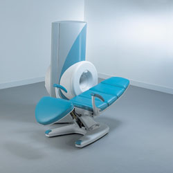
From ONI Medical Systems, Inc.;
MSK-Extremeâ„¢ MRI system is a dedicated high field extremity imaging device, designed to provide orthopedic surgeons and other physicians with detailed diagnostic images of the foot, ankle, knee, hand, wrist and elbow, all with the clinical confidence and advantages derived from high field, whole body MRI units. The light weight (less than 650 kg) of the OrthOne System performs rapid patient studies, is easy to operate, has a patient friendly open environment and can be installed in a practice office or hospital, all at a cost similar to a low field extremity machine.
New features include a more powerful operating system that offers increased scan speed as well as a 160-mm knee coil with higher signal to noise ratio, and the option of a CD burner.
Device Information and Specification 16 cm knee, 18 cm lower extremity;; 12.3 cm upper extremity, additional high resolution v-SPEC Coils: 80 mm, 100 mm, or 145 mm. SE, FSE, GE2D, GE3D, Inversion recovery (IR), Driven Equilibrium, Fat Saturation (FS), STIR, MT, PD, Flow Compensation (FC), RF spoiling, MTE, No Phase Wrap (NPW) IMAGING MODES Scout, single, multislice, volume 2D less than 200 msec/image X/Y: 64-512; 2 pixel steps 4,096 grey lvls; 256 lvls in 3D POWER REQUIREMENTS 115VAC, 1phase, 20A; 208VAC, 3 phase, 30A COOLING SYSTEM TYPE LHe with 2 stage cold head 1.25m radial x 1.8m axial | |  | | | |  Further Reading: Further Reading: | Basics:
|
|
| |
|  |  | Searchterm 'Pixel' was also found in the following services: | | | | |
|  |  |
| |
|
(WFS) The water fat shift defines the frequency bandwidth resulting in pixel shift due to the water/fat spectral separation. WFS decreases or increases to a specified number of pixels in mm. The amount of WFS is proportional to the main magnetic field.
See Bandwidth. | | | |  | |
• View the DATABASE results for 'Water Fat Shift' (7).
| | | | |  Further Reading: Further Reading: | Basics:
|
|
| |
|  | |  |  |
|  | |
|  | | |
|
| |
 | Look
Ups |
| |