Magnetic Resonance - Technology Information Portal Welcome to MRI Technology
Info
  Sheets

Out-
      side


 'Passive Shielding' 
SEARCH FOR    
 
  2 3 5 A B C D E F G H I J K L M N O P Q R S T U V W X Y Z
Result : Searchterm 'Passive Shielding' found in 1 term [] and 2 definitions [], (+ 5 Boolean[] results
1 - 5 (of 8)     next
Result Pages : [1]  [2]
MRI Resources 
DICOM - Mass Spectrometry - Absorption and Emission - Safety pool - Jobs - Process Analysis
 
Passive Shielding
 
Magnetic shielding through the use of high permeability material. The iron provides a return path for the stray field lines of magnetic flux and so significantly decreases the flux away from the magnet.
Passive shielding (see also Faraday cage) significantly eases the problems of siting a MR imager in a confined space. Ferromagnetic objects are less prone to being attracted to the magnet, ancillary electronic equipment, credit cards and computer disks can be brought closer to the magnet and the MRI safety limit for pacemaker wearers (the 5 gauss line = 0.5 mT) is reduced from, typically, 10 m to 2 m from the magnet. A passive shield for a whole-body MRI magnet weights many tons. An alternative method of controlling stray field is active shielding.

See also Active Shielding, Magnetic Shielding, Self Shielding and Room Shielding.
spacer
 
• Share the entry 'Passive Shielding':  Facebook  Twitter  LinkedIn  
 
• Related Searches:
    • Self Shielding
    • Faraday Shield
    • Magnetic Shielding
    • MRI Safety
    • Active Shielding
 
Further Reading:
  Basics:
Faraday's Law
   by hyperphysics.phy-astr.gsu.edu    
  News & More:
Magnetic Sensitivity of MRI Systems to External Iron: The Design Process
   by www.integratedsoft.com    
MRI Resources 
Safety Products - Developers - Process Analysis - Calculation - Movies - DICOM
 
Self Shielding
 
Magnetic shielding by attaching a high permeability yoke to the magnet (passive shielding) or by incorporating additional magnetic field-generating coils designed to reduce the external field (active shielding).

See also Magnetic shielding, Active Shielding and Passive Shielding.
spacer

• View the DATABASE results for 'Self Shielding' (3).Open this link in a new window

MRI Resources 
Safety pool - Musculoskeletal and Joint MRI - MRI Technician and Technologist Jobs - Intraoperative MRI - Mobile MRI - Case Studies
 
Magnetic Shielding
 
Means to confine the region of strong magnetic field surrounding a magnet; most commonly the use of material with high permeability (passive shielding) or by employing secondary counteracting coils outside of the primary coils (active shielding). The high permeability material can be employed in the form of a yoke immediately surrounding the magnet (self-shielding) or installed in the walls of a room as full or partial room-shielding. Unlike shielding ionizing radiation, for example, magnetic shielding can only be accomplished by forcing the unavoidable magnetic return flux through more confined areas or structures, not by absorbing it.

See also Radio Frequency Shielding Radio Frequency Shielding, and Faraday cage.

See also the related poll result: 'Most outages of your scanning system are caused by failure of'
spacer

• View the DATABASE results for 'Magnetic Shielding' (11).Open this link in a new window

 
Further Reading:
  Basics:
Faraday's Law
   by hyperphysics.phy-astr.gsu.edu    
  News & More:
Magnetic Sensitivity of MRI Systems to External Iron: The Design Process
   by www.integratedsoft.com    
MRI Resources 
Societies - PACS - RIS - Veterinary MRI - Chemistry - Stimulator pool
 
C-SCAN™InfoSheet: - Devices -
Intro, 
Types of Magnets, 
Overview, 
etc.MRI Resource Directory:
 - Devices -
 
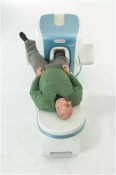
www.gehealthcare.com/usen/mr/cscan/index.html Manufactured by Esaote S.p.A.; compact in-office MRI system, fits in a 90 ft2 (8.4 m2) space and requires no shielding or special power. This low field MRI magnet is optimized for orthopedic use and imaging of the extremities.
The C-SCAN™ is developed from the ARTOSCAN™ - M with a new computer platform, and is also known as Artoscan C.
Esaote North America and Hologic Inc. are the U.S. distributors of this MRI device.
Device Information and Specification
CLINICAL APPLICATION
Dedicated extremity
CONFIGURATION
Closed
Linear and dual phased array, knee, ankle, wrist (6 total) coils
PULSE SEQUENCES
SE, GE, IR, STIR, FSE, 3D CE, GE-STIR, 3D GE, ME, TME, HSE
IMAGING MODES
Single, multislice, volume study, fast scan, multi slab
TR
12 - 5,000 msec
TE
5 - 220 msec
SINGLE SLICE
0.8 sec/image
MULTISLICE
0.8 sec/image
FOV
10 - 20 cm
2D: 2 mm - 10 mm;
3D: 0.6 mm - 10 mm
512 x 512
MEASURING MATRIX
256 x 256 maximum
PIXEL INTENSITY
4,096 gray lvls, 256 lvls in 3D
MAGNET TYPE
Permanent
BORE DIAMETER
or W x H
33.6 x 16 cm
MAGNET WEIGHT
960 kg
H*W*D
124 x 76 x 60 cm
POWER REQUIREMENTS
100/110/200/220/230/240
STRENGTH
10 mT/m
5-GAUSS FRINGE FIELD, radial/axial
28 cm/60 cm
Passive
spacer

• View the DATABASE results for 'C-SCAN™' (4).Open this link in a new window

MRI Resources 
IR - Devices - Hospitals - Contrast Enhanced MRI - DICOM - Non-English
 
O-SCAN™InfoSheet: - Devices -
Intro, 
Types of Magnets, 
Overview, 
etc.
 
www.fonar.com/standup.htm www.fonar.com/standup.htm O-scan is manufactured and distributed by Esaote SpA
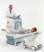
O-scan is a compact, dedicated extremity MRI system designed for easy installation and high throughput. The complete system fits in a 9' x 10' room, doesn't need for RF or magnetic shielding and it plugs in the wall. The 0.31T permanent magnet along with dual phased array RF coils, and advanced imaging protocols provide outstanding image quality and fast 25 minute complete examinations.
Esaote North America is the exclusive distributor of the O-scan system in the USA.
Device Information and Specification
CLINICAL APPLICATION
Dedicated Extremity
CONFIGURATION
Closed
Dual phased array knee, hand, foot//ankle/elbow
PULSE SEQUENCES
SE, HSE, HFE, GE, 2dGE, ME, IR, STIR, Stir T2, GESTIR, TSE, TME, FSE STIR, FSE (T1, T2), X-Bone, Turbo 3DT1, 3D SHARC, 3D SST1, 3D SST2
IMAGING MODES
2D, 3D multi-plane, half echo, half scan, real time
TR
10 - 10,000 msec.
TE
6 - 220 msec.
SINGLE SLICE
0.1 sec.
MULTI SLICE
0.1 sec.
14 cm
2D: 2mm - 10 mm, 3D: 0.6 - 10 mm
MEASURING MATRIX
512 x 512 max.
PIXEL INTENSITY
4,096 grey levels
MAGNET TYPE
Permanent - NdFeB
MAGNET WEIGHT
2,733 lbs
POWER REQUIREMENTS
100/110/200/220/230/240
STRENGTH
20 mT/m
5 GAUSS FRINGE FIELD, radial/axial
67 cm / 75 cm
passive
spacer
MRI Resources 
Contrast Agents - Homepages - Equipment - Service and Support - Non-English - Services and Supplies
 
     1 - 5 (of 8)     next
Result Pages : [1]  [2]
 Random Page
 
Share This Page
FacebookTwitterLinkedIn

MR-TIP    
Community   
User
Pass
Forgot your UserID/Password ?    


MRI is trending to low field magnets :
reduced costs will lead to this change 
AI will close the gap to high field 
only in remote areas 
is only temporary 
never 

Look
      Ups


MR-TIP.com uses cookies! By browsing MR-TIP.com, you agree to our use of cookies.

Magnetic Resonance - Technology Information Portal
Member of SoftWays' Medical Imaging Group - MR-TIP • Radiology-TIP • Medical-Ultrasound-Imaging • 
Copyright © 2003 - 2024 SoftWays. All rights reserved. [ 24 April 2024]
Terms of Use | Privacy Policy | Advertising
 [last update: 2024-02-26 03:41:00]