Magnetic Resonance - Technology Information Portal Welcome to MRI Technology
Info
  Sheets

Out-
      side


 'Navigator Technique' 
SEARCH FOR    
 
  2 3 5 A B C D E F G H I J K L M N O P Q R S T U V W X Y Z
Result : Searchterm 'Navigator Technique' found in 1 term [] and 3 definitions [], (+ 1 Boolean[] results
1 - 5 (of 5)     
Result Pages : [1]
MRI Resources 
Service and Support - Open Directory Project - Abdominal Imaging - Education pool - Crystallography - Coils
 
Navigator Technique
 
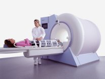
The navigator technique measures with an additional quick MR prepulse the position, of e.g. the diaphragm before data collecting. Similar respiratory conditions of the patient can be identified and used to synchronize image data acquisition so that respiration induced image blurring is minimized by either respiratory ordered phase encoding or respiratory gating.
The prepulse sequence images a small area perpendicular to the structure, which is moving. The contrast of the interface between the diaphragm and the lung should be high to permit easy automatic detection. After data acquisition, the position of the interface is automatically recorded and imaging data are only accepted when the position of the interface falls within a range of prespecified values.
This technique has the advantage of greater accuracy than other respiratory gating (therefore used for coronary angiography) and has no need for additional sensing MRI equipment, as the MR system itself provides it.
spacer
 
• Share the entry 'Navigator Technique':  Facebook  Twitter  LinkedIn  
 
• Related Searches:
    • Heart MRI
    • Breath Hold Imaging
    • Cardiac MRI
    • Cardiac Gating
    • Respiratory Compensation
MRI Resources 
Devices - Coils - Education pool - Chemistry - Nerve Stimulator - MR Guided Interventions
 
Coronary AngiographyMRI Resource Directory:
 - Cardiovascular Imaging -
 
(MRI-CA, MRCA) The noninvasive imaging of the coronary arteries using magnetic resonance imaging of the heart.
For cardiac MRI-CA, high performance machines are necessary with minimum 40mT/m and 300μsec slew rate.
2D and 3D acquisition are used for fast gradient echo sequences with techniques for minimizing cardiac and respiratory motion and suppressing the high signal of pericardial fat. The optimal sequences seem to be trueFISP, Balanced FFE or FIESTA with SMASH and SENSE techniques. Respiratory motion is minimized for 3D acquisitions by using respiratory gating, especially using navigator echoes (Navigator Technique) to track diaphragmatic and cardiac movement. Optimization of MR technique can provide mapping of long segments of the coronary arteries.
Blood pool agents are being applied to improve the reliability of coronary MR angiography. The major current clinical indication is the identification of coronary artery anomalies because the diagnostic accuracy's for identifying haemodynamically significant stenoses are variable depending of the image quality.

See also Magnetic Resonance Angiography, and Cardiac MRI.
spacer

• View the DATABASE results for 'Coronary Angiography' (7).Open this link in a new window

 
Further Reading:
  Basics:
Role of Magnetic Resonance Imaging in Visualizing Coronary Arteries
Monday, 2 August 2004   by www.clinmedres.org    
  News & More:
Graphic illustration
Tuesday, 12 February 2008   by www.theengineer.co.uk    
MRI Resources 
Most Wanted - Claustrophobia - Safety pool - MRCP - Collections - DICOM
 
Fast Imaging Employing Steady State AcquisitionInfoSheet: - Sequences - 
Intro, 
Overview, 
Types of, 
etc.
 
(FIESTA) The fast imaging employing steady state acquisition sequence provides images of fluid filled structures with very short acquisition times. The FIESTA sequence uses the T2 steady state contrast mechanism to provide high SNR images with strong signal from fluid tissues while suppressing background tissue for contrast and anatomic detail of small structures. In addition, the ultra short TR and TE enable extremely short acquisition times - shorter than FSE - and the images can be post processed using MIP, volume rendering, or 3D navigator techniques.

See Steady State Free Precession.
spacer
MRI Resources 
Claustrophobia - Libraries - - Resources - General - Supplies
 
MAGNETOM Sonata™InfoSheet: - Devices -
Intro, 
Types of Magnets, 
Overview, 
etc.MRI Resource Directory:
 - Devices -
 
www.med.siemens.com/med/d/gg/mr/products/sonata.html From Siemens Medical Systems;
while older navigator techniques take up to 40 minutes to create, the high performance of the MAGNETOM Sonata system enables 'complete examinations in less than 15 minutes'. It creates a new standard of diagnostic confidence and moves Cardiac MR from the research setting into routine clinical practice.
Device Information and Specification
CLINICAL APPLICATION
Whole body
CONFIGURATION
Compact
Yes/SVS or CSI opt.
SYNCHRONIZATION
ECG/peripheral, respiratory gating
PULSE SEQUENCES
GRE, IR, FIR, STIR, TrueIR/FISP, FSE, FLAIR, MT, SS-FSE, MT-SE, MTC, MSE, EPI, 3D DESS//CISS/PSIF, GMR
IMAGING MODES
Single, multislice, volume study, multi angle, multi oblique
TR
1.5 msec at 256matrix
TE
0.58 msec at 256matrix
SINGLE/MULTI SLICE
178 images/sec at 256 x 256 at 100% FOV
0.5 cm - 40 cm
Min 2D/3D: 0.1/0.05 mm
1024 x 1024 full screen display
MEASURING MATRIX
64 x 64 to 1024 x 1024
PIXEL INTENSITY
10 micrometer in plane
60 cm
MAGNET WEIGHT
4050kg, 5500kg in operation
H*W*D
236 x 215 x 160 cm
POWER REQUIREMENTS
380/400/420/440/480 V
COOLING SYSTEM TYPE
Single cryogen, 2 stage refrig.
Liquid He
STRENGTH
40/70 mT/m
5-GAUSS FRINGE FIELD
2.5 m / 4.0 m
Passive, act.; 1st order std./2nd opt.
spacer

• View the DATABASE results for 'MAGNETOM Sonata™' (2).Open this link in a new window

MRI Resources 
Absorption and Emission - RIS - Abdominal Imaging - Contrast Agents - Process Analysis - Online Books
 
Diffusion Weighted SequenceInfoSheet: - Sequences - 
Intro, 
Overview, 
Types of, 
etc.MRI Resource Directory:
 - Diffusion Weighted Imaging -
 
Diffusion weighted imaging can be performed similar to the phase contrast angiography sequence. The gradients must be increased in amplitude to depict the much slower motions of molecular diffusion in the body.
While a T1 weighted MRI pulse sequence is diffusion sensitive, a quantitative diffusion pulse sequence was introduced by Steijskal and Tanner. Its characteristic features are two strong symmetrical gradient lobes placed on either side of the 180° refocusing pulse in a spin echo sequence. These symmetrical gradient lobes have the sole purpose of enhancing dephasing of spins, thereby accelerating intravoxel incoherent motion (IVIM) signal loss.
Dephasing is proportional to the square of the time (diffusion time) during which the gradients are switched on and the strength of the applied gradient field. Therefore, the use of high field gradient systems with faster and more sensitive sequences, make diffusion weighting more feasible.
Areas in which the protons diffuse rapidly (swollen cells in early stroke, less restriction to diffusion) will show an increased signal when the echo is measured relative to areas in which diffusion is restricted. For increased accuracy of diffusion measurement and image enhancement, useful motion correction techniques such as navigator echo and other methods should be used. In addition to this, applying the b-value calculated by the strength and duration of motion probing gradients with a high rate of accuracy is very important.

See also Apparent Diffusion Coefficient, ADC Map, Lattice Index Map.
spacer

• View the DATABASE results for 'Diffusion Weighted Sequence' (6).Open this link in a new window

 
Further Reading:
  Basics:
Diffusion-Weighted Imaging
   by spinwarp.ucsd.edu    
A Comparison of Methods for High-Spatial-Resolution Diffusion-weighted Imaging in Breast MRI
Tuesday, 25 August 2020   by pubs.rsna.org    
Diffusion Imaging: From Basic Physics to Practical Imaging
1999   by ej.rsna.org    
  News & More:
DWI-MRI helps breast cancer patients' chemotherapy response
Friday, 20 January 2023   by www.auntminnieeurope.com    
Effect of gadolinium-based contrast agent on breast diffusion-tensor imaging
Thursday, 6 August 2020   by www.eurekalert.org    
Hopkins researchers use diffusion MRI technique to monitor ultrasound uterine fibroid treatment
Monday, 8 August 2005   by www.eurekalert.org    
Diffusion-weighted MRI sensitive for metastasis in pelvic lymph nodes
Sunday, 15 June 2014   by www.2minutemedicine.com    
EVALUATION OF HUMAN STROKE BY MR IMAGING
2000
MRI Resources 
Claustrophobia - Societies - Safety Training - Breast Implant - MRI Physics - General
 
     1 - 5 (of 5)     
Result Pages : [1]
 Random Page
 
Share This Page
FacebookTwitterLinkedIn

MR-TIP    
Community   
User
Pass
Forgot your UserID/Password ?    


MRI is trending to low field magnets :
reduced costs will lead to this change 
AI will close the gap to high field 
only in remote areas 
is only temporary 
never 

Look
      Ups


MR-TIP.com uses cookies! By browsing MR-TIP.com, you agree to our use of cookies.

Magnetic Resonance - Technology Information Portal
Member of SoftWays' Medical Imaging Group - MR-TIP • Radiology-TIP • Medical-Ultrasound-Imaging • 
Copyright © 2003 - 2024 SoftWays. All rights reserved. [ 20 April 2024]
Terms of Use | Privacy Policy | Advertising
 [last update: 2024-02-26 03:41:00]