Magnetic Resonance - Technology Information Portal Welcome to MRI Technology
Info
  Sheets

Out-
      side


 'Magnetic Resonance Tomography' 
SEARCH FOR    
 
  2 3 5 A B C D E F G H I J K L M N O P Q R S T U V W X Y Z
Result : Searchterm 'Magnetic Resonance Tomography' found in 1 term [] and 2 definitions [], (+ 10 Boolean[] results
previous     6 - 10 (of 13)     next
Result Pages : [1]  [2 3]
Searchterm 'Magnetic Resonance Tomography' was also found in the following services: 
spacer
News  (39)  Resources  (26)  
 
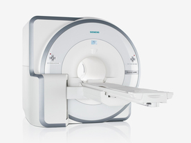
Biograph mMR
 
www.healthcare.siemens.com/magnetic-resonance-imaging/mr-pet-scanner/biograph-mmr FDA cleared and CE Mark 2011.
The Biograph mMR has a fully-integrated design for simultaneous PET/MRI imaging. The dedicated hardware includes solid-state, avalanche photodiode PET detector and adapted, PET-compatible MR coils.
The possibility of truly simultaneous operation allows the acquisition of several magnetic resonance imaging (MRI) sequences during the positron emission tomography (PET) scan, without increasing the examination time.
See also Hybrid Imaging.
Device Information and Specification
CLINICAL APPLICATION
Whole Body
CONFIGURATION
Simultaneous PET/MRI
3 Tesla
Full range of MRI coils
CHANNELS
up to 32
PET ACQUISITION MODES
3D
MRI ACQUISITION MODES
2D/3D, spectroscopy;; iPAT, GRAPPA (k-space), noncontrast angiography, radial motion compensation
PET EFFECTIVE AXIAL FOV
26 cm (typical overlap 23%)
MRI FOV
A-P 45, R-L 50, H-F 50 cm
PET RING DIAMETER
65.6 cm
PATIENT SCAN RANGE
199 cm
HORIZONTAL SPEED
200 mmsec
TABLE CAPACITY
200 kg
PET DETECTOR
Solid state, 4032 avalanche photo diodes
DETECTOR SCINTILLATION MATERIAL
LSO, 28672 crystals
CRYSTAL SIZE
4 x 4 x 20 mm
MAGNET WEIGHT
9000 kg
DIMENSION H*W*D (gantry included)
335 x 230 x 242 cm (finshed covers)
5-GAUSS FRINGE FIELD
2.6 m / 4.6 m
CRYOGEN USE
Zero boil off rate, approx. 10 years
COOLING SYSTEM
PET system: water; MRI system: water
up to 200 T/m/s
MAX. AMPLITUDE
45 mT/m
Aautomatic, patient specific shim; active shim 3 linear and 5 non-linear channels (seond order)
POWER REQUIREMENTS
380 / 400 / 420 / 440 / 460 / 480 V, 3-phase + ground; Total system 110kW
spacer
 
Further Reading:
  Basics:
Performance Measurements of the Siemens mMR Integrated Whole-Body PET/MR Scanner
Friday, 11 November 2011   by jnm.snmjournals.org    
Global Trends in Hybrid Imaging
Monday, 1 November 2010   by pubs.rsna.org    
  News & More:
Positron Emission Tomographic Imaging in Stroke
Monday, 28 December 2015   by www.ncbi.nlm.nih.gov    
A world's first in imaging – integrated whole-body molecular MR system available for clinical use testing
Thursday, 18 November 2010   by www.siemens.com    
FDA Clears New System to Perform Simultaneous PET, MRI Scans
Friday, 10 June 2011   by www.prnewswire.com    
Frost & Sullivan Award Underlines Siemens Healthcare's Contribution to New Product Innovation in the North American Medical Imaging Market
Wednesday, 13 July 2011   by multivu.prnewswire.com    
Searchterm 'Magnetic Resonance Tomography' was also found in the following service: 
spacer
Radiology  (1) Open this link in a new window
Lung ImagingMRI Resource Directory:
 - Lung Imaging -
 
Lung imaging is furthermore a challenge in MRI because of the predominance of air within the lungs and associated susceptibility issues as well as low signal to noise of the inflated lung parenchyma. Cardiac and respiratory triggered or breath hold sequences allow diagnostic imaging, however a comparable image quality with computed tomography is still difficult to achieve.
Assumptions for lung MRI:
•
Low signal to noise ratio of the inherently low lung proton density.
•
Cardiac and respiratory motion artifacts.
•
Magnetic susceptibility effects of large magnetic field gradients.
•
Very short transverse relaxation times and significant diffusion yielding short T2 (30-70 msec), short T2* (1-3 msec), and additional long T1 relaxation times (1300-1500 msec).
•
The extreme short T2 values are responsible for a fast signal decay during a single shot readout, resulting in blurring.

The current trends in MRI are the use of new imaging technologies and increasingly powerful magnetic fields. Among these technologies are parallel imaging techniques as well as ventilation agents like hyperpolarized helium for the use as an inert inhalational contrast agent to study lung ventilation properties. With hyperpolarized gases clear images of the lungs can be obtained without using a large magnetic field (see also back projection imaging). Single shot sequences (e.g. TSE or Half Fourier Acquisition Single Shot Turbo Spin Echo HASTE) used in lung MR imaging benefits from parallel imaging techniques due to reduced relaxation time effects during the echo train and therefore reduced image blurring as well as reduced motion artifacts.
In the future, more effective contrast agents may provide an alternative solution to the need for high field MRI. Dynamic contrast enhanced MRI perfusion has demonstrated a potential in the diagnosis of pulmonary embolism or to characterize lung cancer and mediastinal tumors. 3D contrast enhanced magnetic resonance angiography of the thoracic vessel.

See also the related poll result: 'MRI will have replaced 50% of x-ray exams by'
 
Images, Movies, Sliders:
 Anatomic Imaging of the Lungs  Open this link in a new window
      

Courtesy of  Robert R. Edelman
 Normal Lung Gd Perfusion MRI  Open this link in a new window
      

Courtesy of  Robert R. Edelman

 MRI Thorax Basal Plane  Open this link in a new window
 
Radiology-tip.comradLung Scintigraphy
spacer

• View the DATABASE results for 'Lung Imaging' (7).Open this link in a new window


• View the NEWS results for 'Lung Imaging' (3).Open this link in a new window.
 
Further Reading:
  Basics:
A safer approach for diagnostic medical imaging
Monday, 29 September 2014   by www.eurekalert.org    
Parallel Lung Imaging(.pdf)
  News & More:
Chest MRI a viable alternative to chest CT in COVID-19 pneumonia follow-up
Monday, 21 September 2020   by www.healthimaging.com    
CT Imaging Features of 2019 Novel Corona virus (2019-nCoV)
Tuesday, 4 February 2020   by pubs.rsna.org    
Polarean Imaging Phase III Trial Results Point to Potential Improvements in Lung Imaging
Wednesday, 29 January 2020   by www.diagnosticimaging.com    
Low Power MRI Helps Image Lungs, Brings Costs Down
Thursday, 10 October 2019   by www.medgadget.com    
Chest MRI Using Multivane-XD, a Novel T2-Weighted Free Breathing MR Sequence
Thursday, 11 July 2019   by www.sciencedirect.co    
Researchers Review Importance of Non-Invasive Imaging in Diagnosis and Management of PAH
Wednesday, 11 March 2015   by lungdiseasenews.com    
New MRI Approach Reveals Bronchiectasis' Key Features Within the Lung
Thursday, 13 November 2014   by lungdiseasenews.com    
MRI techniques improve pulmonary embolism detection
Monday, 19 March 2012   by medicalxpress.com    
  News & More:
Partnership with VIDA to streamline adoption of advanced MRI of the lungs
Monday, 11 September 2023   by www.itnonline.com    
MRI Resources 
Fluorescence - Manufacturers - Libraries - Breast Implant - Homepages - Veterinary MRI
 
Molecular Imaging
 
Molecular Imaging is a new diagnostic discipline to visualize biological processes.
Molecular magnetic resonance imaging (mMRI) offers the potential to image tissues at the cellular and subcellular level. Targeted MR contrast agents enhance the diagnostic specificity and range of molecular magnetic resonance imaging.
Other modalities that can be used for noninvasive molecular imaging:
•
Ultrasound;
•
optical imaging;
•
positron emission tomography (PET);
•
single photon emission computed tomography (SPECT).


See also Nanoparticle, Monocrystalline Iron Oxide Nanoparticle, Polycrystalline Iron Oxide Nanoparticles, Liposomes, Monoclonal Antibodies, Bimodal Imaging, Tumor Specific Agents, and Intracellular Contrast Agents.
spacer

• View the DATABASE results for 'Molecular Imaging' (10).Open this link in a new window


• View the NEWS results for 'Molecular Imaging' (28).Open this link in a new window.
 
Further Reading:
  Basics:
Multimodal Nanoparticles for Quantitative Imaging(.pdf)
Tuesday, 13 December 2011   by alexandria.tue.nl    
Molecular Magnetic Resonance Imaging(.pdf)
2005   by www.medical.siemens.com    
  News & More:
Smarter MRI diagnosis with nano MRI lamp
Monday, 6 February 2017   by www.eurekalert.org    
Molecular MRI technique gives early indication of cancer treatment effectiveness
Monday, 11 April 2016   by www.healthimaging.com    
Molecular imaging and radiochemistry: the importance of instrumentation. An interview with Professor Bjorn Wangler
Thursday, 4 February 2016   by www.news-medical.net    
Positron Emission Tomographic Imaging in Stroke
Monday, 28 December 2015   by www.ncbi.nlm.nih.gov    
Potential and Limitations of Oxygen-17 MR Perfusion Measurements
Monday, 1 March 2004   by www.case.edu    
Searchterm 'Magnetic Resonance Tomography' was also found in the following services: 
spacer
News  (39)  Resources  (26)  
 
Cervical Spine MRI
 
Cervical spine MRI is a suitable tool in the assessment of all cervical spine (vertebrae C1 - C7) segments (computed tomography (CT) images may be unsatisfactory close to the thoracic spine due to shoulder artifacts). The cervical spine is particularly susceptible to degenerative problems caused by the complex anatomy and its large range of motion.
Advantages of magnetic resonance imaging MRI are the high soft tissue contrast (particularly important in diagnostics of the spinal cord), the ability to display the entire spine in sagittal views and the capacity of 3D visualization. Magnetic resonance myelography is a useful supplement to conventional MRI examinations in the investigation of cervical stenosis. Myelographic sequences result in MR images with high contrast that are similar in appearance to conventional myelograms. Additionally, open MRI studies provide the possibility of weight-bearing MRI scan to evaluate structural positional and kinetic changes of the cervical spine.
Indications of cervical spine MRI scans include the assessment of soft disc herniations, suspicion of disc hernia recurrence after operation, cervical spondylosis, osteophytes, joint arthrosis, spinal canal lesions (tumors, multiple sclerosis, etc.), bone diseases (infection, inflammation, tumoral infiltration) and paravertebral spaces.
State-of-the-art phased array spine coils and high performance MRI machines provide high image quality and short scan time. Imaging protocols for the cervical spine includes sagittal T1 weighted and T2 weighted sequences with 3-4 mm slice thickness and axial slices; usually contiguous from C2 through T1. Additionally, T2 fat suppressed and T1 post contrast images are often useful in spine imaging.

See also Lumbar Spine MRI.
spacer

• View the DATABASE results for 'Cervical Spine MRI' (2).Open this link in a new window


• View the NEWS results for 'Cervical Spine MRI' (1).Open this link in a new window.
 
Further Reading:
  News & More:
Ultrafast MRI protocol reduces scan time by 10 minutes for cervical imaging
Monday, 26 September 2022   by healthimaging.com    
In Vivo 3-D Cervical Spine Kinematics Demonstrated
Thursday, 19 May 2011   by www.doctorslounge.com    
MRI Images at a 45-Degree Angle Through The Cervic al Neural Forami na:A Technique For Improved Visualization(.pdf)
2006   by www.painphysicianjournal.com    
Searchterm 'Magnetic Resonance Tomography' was also found in the following service: 
spacer
Radiology  (1) Open this link in a new window
Elscint Ltd.MRI Resource Directory:
 - Manufacturers -
 
Founded in 1969, Elscint is headquartered in Haifa, Israel. Elscint developed advanced computerized imaging systems in Computed Tomography (CT), Magnetic Resonance Imaging (MRI), Nuclear Medicine (NM) and Mammography (MAM) for international markets.
In November 1998, General Electric Medical Systems (GEMS) acquired the Nuclear Medicine and MRI divisions of Elscint, including an unique MRI gradient system concept and technology (twin gradient system).
Elscint Ltd. signs definitive agreement to sell its Nuclear Medicine and Magnetic Resonance Imaging Businesses for $100 Million. Elscint's shareholders approved the sale of its NM, and MRI division to GE (now GE Healthcare). Picker International acquires the Computed Tomography Division of Elscint Ltd. in the same year.

See also Marconi Medical Systems.
spacer
 
Further Reading:
  Basics:
Elscint's Shareholders Approved The Sale of its NM, MRI and CT Division To GE and Picker
Monday, 23 November 1998   by www.prnewswire.com    
  News & More:
Twin Gradient Technology - Potential Advantages For Diffusion Weighted MRI(.pdf)
   by www.paulrharvey.co.uk    
MRI Resources 
Spine MRI - Devices - Contrast Agents - Knee MRI - Shoulder MRI - Mobile MRI Rental
 
previous      6 - 10 (of 13)     next
Result Pages : [1]  [2 3]
 Random Page
 
Share This Page
FacebookTwitterLinkedIn

MR-TIP    
Community   
User
Pass
Forgot your UserID/Password ?    


MRI is trending to low field magnets :
reduced costs will lead to this change 
AI will close the gap to high field 
only in remote areas 
is only temporary 
never 

Look
      Ups


MR-TIP.com uses cookies! By browsing MR-TIP.com, you agree to our use of cookies.

Magnetic Resonance - Technology Information Portal
Member of SoftWays' Medical Imaging Group - MR-TIP • Radiology-TIP • Medical-Ultrasound-Imaging • 
Copyright © 2003 - 2024 SoftWays. All rights reserved. [ 24 April 2024]
Terms of Use | Privacy Policy | Advertising
 [last update: 2024-02-26 03:41:00]