| Info
Sheets |
| | | | | | | | | | | | | | | | | | | | | | | | |
 | Out-
side |
| | | | |
|
| | | | | |  | Searchterm 'Magnet' was also found in the following services: | | | | |
|  |  |
| |
|
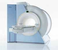
From Siemens Medical Systems;
the MAGNETOM Rhapsody™. This open MRI system offers the proven image
quality of 1.0 Tesla. In addition to the resulting broad range of applications, the open magnet of the high field system MAGNETOM Rhapsodyâ„¢ facilitates examination of claustrophobic and pediatric patients. And the system allows for expanded interventional applications.
Device Information and Specification CLINICAL APPLICATION Whole body GRE, IR, FIR, STIR, TrueIR/FISP, FSE, FLAIR, MT, SS-FSE, MT-SE, MTC, MSE, EPI, GMR, fat/water sat./exc. IMAGING MODES Single, multislice, volume study, multi angle, multi oblique1024 x 1024 full screen display POWER REQUIREMENTS 380/400/420/440/480 V | | | | | |
| | | Searchterm 'Magnet' was also found in the following services: | | | | |
| | |
| |
|
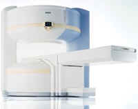
Device Information and Specification CLINICAL APPLICATION Whole body GRE, IR, FIR, STIR, TrueIR/FISP, FSE, FLAIR, MT, SS-FSE, MT-SE, MTC, MSE, EPI, GMR, fat/water sat./exc. IMAGING MODES Single, multislice, volume study, multi angle, multi obliqueTR 2.4 msec std.; 2.0 opt.; 1.8 w/30 mT/m at 256matrix TE 1.1 msec std.; 0.9 opt.; 0.78 w/30 mT/m at 256matrix 178 images/sec at 256 x 256 at 100% FOV1024 x 1024 full screen display 21 micrometer in plane, 11 micrometer optional 4050kg, 5500kg in operation H*W*D 236 x 215 x 160 cm w/covers POWER REQUIREMENTS 380/400/420/440/480 V STRENGTH 20/35 mT/m standard, 30/52 opt. Passive, act.; 1st order std./2nd opt. | | | |
• View the DATABASE results for 'MAGNETOM Symphony™' (2).
| | | | |  Further Reading: Further Reading: | Basics:
|
|
| |
| | | | | |
| |
|
The region surrounding a magnet and exhibiting a magnetic field strength, which is significantly higher than the earth's magnetic field (typically 0.05-0.1 mT, depending on geographical location).
Initially the most magnets had very extensive fringe fields. Magnets with iron have reduced the fringe field substantially (passively shielded magnets). At least, adding appropriate additional superconducting coils to superconducting magnets has resulted in a drastic reduction of the extent of the fringe fields (actively shielded magnets).
Due to the physical properties of magnetic fields, the magnetic flux, which penetrates the useful volume of the magnet will return through the surroundings of the magnet to form closed field lines. Depending on the magnet construction, the returning flux will penetrate large open spaces (unshielded magnets) or will be confined largely to iron yokes or through secondary coils (shielded magnets).
Fringe fields constitute one of the major hazards of MR scanners as these fields acting over extended distances outside the magnet produce strong attractive forces upon magnetic objects. These can thus 'fly' into the magnet when loose nearby acting like projectiles. Fringe fields also exert unwanted forces on metallic implants in patients. | | | |
• View the DATABASE results for 'Magnetic Fringe Field' (3).
| | | | | | Further Reading: | Basics:
|
|
| |
| | | Searchterm 'Magnet' was also found in the following services: | | | | |
| | |
| |
|
| | | | | | | Further Reading: | News & More:
|
|
| |
| | | Searchterm 'Magnet' was also found in the following services: | | | | |
| | |
| |
|
(MT) Magnetization Transfer was accidentally discovered by Wolff and Balaban in 1989. Conventional MRI is based on the differences in T1, T2 and the proton density (water content and the mobility of water molecules) in tissue; it relies primarily on free (bulk) water protons. The T2 relaxation times are greater than 10 ms and detectable. The T2 relaxation times of protons associated with macromolecules are less then 1 ms and not detectable in MRI.
Magnetization Transfer Imaging (MTI) is based on the magnetization interaction (through dipolar and/or chemical exchange) between bulk water protons and macromolecular protons. By applying an off resonance radio frequency pulse to the macromolecular protons, the saturation of these protons is then transferred to the bulk water protons. The result is a decrease in signal (the net magnetization of visible protons is reduced), depending on the magnitude of MT between tissue macromolecules and bulk water. With MTI, the presence or absence of macromolecules (e.g. in membranes, brain tissue) can be seen.
The magnetization transfer ratio (MTR) is the difference in signal intensity with or without MT.
See also Magnetization Transfer Contrast. | | | |
• View the DATABASE results for 'Magnetization Transfer' (7).
| | | | | | Further Reading: | | Basics:
|
|
News & More:
| |
| |
| | | | |
| | |
| | | |
|
| |
 | Look
Ups |
| |