Contrast is the relative difference of signal intensities in two adjacent regions of an image.
Due to the
T1 and
T2 relaxation properties in
magnetic resonance imaging, differentiation between various tissues in the body is possible. Tissue contrast is affected by not only the
T1 and
T2 values of specific tissues, but also the differences in the
magnetic field strength, temperature changes, and many other factors. Good tissue contrast relies on optimal selection of appropriate pulse
sequences (
spin echo,
inversion recovery,
gradient echo, turbo
sequences and
slice profile).
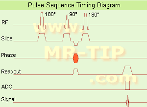
Important
pulse sequence parameters are TR (
repetition time), TE (time to
echo or
echo time), TI (time for
inversion or
inversion time) and
flip angle. They are associated with such parameters as
proton density and
T1 or
T2 relaxation times. The values of these parameters are influenced differently by different tissues and by healthy and diseased sections of the same tissue.
For the
T1 weighting it is important to select a correct TR or TI.
T2 weighted images depend on a correct choice of the TE. Tissues vary in their
T1 and
T2 times, which are manipulated in
MRI by selection of TR, TI, and TE, respectively. Flip angles mainly affect the strength of the signal measured, but also affect the TR/TI/TE parameters.
Conditions necessary to produce different weighted images:
T1 Weighted Image: TR value equal or less than the tissue specific
T1 time - TE value less than the tissue specific
T2 time.
T2 Weighted Image: TR value much greater than the tissue specific
T1 time - TE value greater or equal than the tissue specific
T2 time.
Proton Density Weighted Image: TR value much greater than the tissue specific
T1 time - TE value less than the tissue specific
T2 time.
See also
Image Contrast Characteristics,
Contrast Reversal,
Contrast Resolution, and
Contrast to Noise Ratio.


 (IR) The