 | Info
Sheets |
| | | | | | | | | | | | | | | | | | | | | | | | |
 | Out-
side |
| | | | |
|
| | | | |
Result : Searchterm 'H1' found in 1 term [ ] and 20 definitions [ ] and 20 definitions [ ] ]
| | 1 - 5 (of 21) nextResult Pages : [1] [2 3 4 5] |  | |  | Searchterm 'H1' was also found in the following services: | | | | |
| |  |
| |
|
| | | | | | • Share the entry 'H1':    | | | | |  Further Reading: Further Reading: | News & More:
|
|
| |
| | | Searchterm 'H1' was also found in the following service: | | | | |
| |  |
| |
|

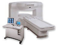
From ONI Medical Systems, Inc.;
MSK-Extreme™ MRI system is a dedicated high field extremity imaging device, designed to provide orthopedic surgeons and other physicians with detailed diagnostic images of the foot, ankle, knee, hand, wrist and elbow, all with the clinical confidence and advantages derived from high field, whole body MRI units. The light weight (less than 650 kg) of the OrthOne System performs rapid patient studies, is easy to operate, has a patient friendly open environment and can be installed in a practice office or hospital, all at a cost similar to a low field extremity machine.
New features include a more powerful operating system that offers increased scan speed as well as a 160-mm knee coil with higher signal to noise ratio, and the option of a CD burner.
Device Information and Specification 16 cm knee, 18 cm lower extremity;; 12.3 cm upper extremity, additional high resolution v-SPEC Coils: 80 mm, 100 mm, or 145 mm. SE, FSE, GE2D, GE3D, Inversion recovery (IR), Driven Equilibrium, Fat Saturation (FS), STIR, MT, PD, Flow Compensation (FC), RF spoiling, MTE, No Phase Wrap (NPW) IMAGING MODES Scout, single, multislice, volume 2D less than 200 msec/image X/Y: 64-512; 2 pixel steps 4,096 grey lvls; 256 lvls in 3D POWER REQUIREMENTS 115VAC, 1phase, 20A; 208VAC, 3 phase, 30A COOLING SYSTEM TYPE LHe with 2 stage cold head 1.25m radial x 1.8m axial | | | | | | | Further Reading: | Basics:
|
|
| |
| | | | | |
| |
|
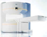
From GE Healthcare;
the New Signa Profile/i is a patient friendly open MRI system that virtually eliminates patient anxiety and claustrophobia, without compromising diagnostic utility.
Device Information and Specification CLINICAL APPLICATION Whole body Integrated transmit body coil, body flex sizes: M, L, XL, quadrature, head coil quadrature, 4 channel neurovascular array, 8 channel CTL array, quad. c- spine, 2 channel shoulder array, extremity coil, 3 channel wrist array, 4 channel breast array, 6, 9, 11 inch general purpose loop coils Standard: SE, IR, 2D/3D GRE and SPGR, Angiography: 2D/3D TOF, 2D/3D phase contrast; 2D/3D FSE, 2D/3D FRFSE, FGRE and FSPGR, SSFP, FLAIR, EPI, optional: 2D/3D Fiesta, fat/water separation, T1 FLAIRIMAGING MODES Localizer, single slice, multislice, volume, fast, POMP, multi slab, cine, slice and frequency zip, extended dynamic range, tailored RF TR 6 to 12000 msec in increments of 1 msec TE 1.3 to 2000 msec in increments of 1 msec 2D: 2.7mm - 20mm 3D: 0.2mm - 5mm 0.08 mm; 0.02 mm optional 10,000 kg w/gradient enclosure POWER REQUIREMENTS 200 - 480, 3-phase COOLING SYSTEM TYPE None required | | | |
• View the DATABASE results for 'Signa Profile™' (2).
| | | | |
| | | Searchterm 'H1' was also found in the following services: | | | | |
| | |
| |
|
[B 1] A conventional symbol for the radio frequency field strength (another symbol historically used, is H1). In MRI, B1 labels the field produced by the radio frequency coil.
The B1 field is often conceived of two vectors rotating in opposite directions, usually in a plane transverse to B0. At the Larmor frequency, the vector rotating in the same direction as the precessing spins will interact strongly with the spins. | | | |
• View the DATABASE results for 'B1' (1346).
| | | | | | Further Reading: | News & More:
|
|
| |
| | | Searchterm 'H1' was also found in the following service: | | | | |
| | |
| |
|
From Siemens Medical Systems;
the MAGNETOM Rhapsody™. This open MRI system offers the proven image
quality of 1.0 Tesla. In addition to the resulting broad range of applications, the open magnet of the high field system MAGNETOM Rhapsodyâ„¢ facilitates examination of claustrophobic and pediatric patients. And the system allows for expanded interventional applications.
Device Information and Specification CLINICAL APPLICATION Whole body GRE, IR, FIR, STIR, TrueIR/FISP, FSE, FLAIR, MT, SS-FSE, MT-SE, MTC, MSE, EPI, GMR, fat/water sat./exc. IMAGING MODES Single, multislice, volume study, multi angle, multi oblique1024 x 1024 full screen display POWER REQUIREMENTS 380/400/420/440/480 V | | | |
• View the DATABASE results for 'MAGNETOM Rhapsody™' (2).
| | | | |
| | | | |
| | |
| | | 1 - 5 (of 21) nextResult Pages : [1] [2 3 4 5] |
| |
|
| |
 | Look
Ups |
| |


