Magnetic Resonance - Technology Information Portal Welcome to MRI Technology
Info
  Sheets

Out-
      side


 'Gradient Echo' 
SEARCH FOR    
 
  2 3 5 A B C D E F G H I J K L M N O P Q R S T U V W X Y Z
Result : Searchterm 'Gradient Echo' found in 20 terms [] and 114 definitions []
previous     11 - 15 (of 134)     next
Result Pages : [1 2 3 4]  [5 6 7 8 9 10 11 12 13 14 15 16 17 18 19 20 ... ]
Searchterm 'Gradient Echo' was also found in the following services: 
spacer
News  (1)  Resources  (2)  Forum  (6)  
 
Refocused Gradient Echo SequenceInfoSheet: - Sequences - 
Intro, 
Overview, 
Types of, 
etc.MRI Resource Directory:
 - Sequences -
 
Refocused GRE sequences use a refocusing gradient in the phase encoding direction during the end module to maximize (refocus) remaining xy- (transverse) magnetization at the time when the next excitation is due, while the other two gradients are, in any case, balanced.
When the next excitation pulse is sent into the system with an opposed phase, it tilts the magnetization in the α direction. As a result the z-magnetization is again partly tilted into the xy-plane, while the remaining xy-magnetization is tilted partly into the z-direction.
Companies use different acronyms to describe certain techniques.

Different terms for these gradient echo pulse sequences
R-GRE Refocused Gradient Echo,
FAST Fourier Acquired Steady State,
FFE Fast Field echo,
FISP Fast Imaging with Steady State Precession,
F-SHORT SHORT Repetition Technique Based on Free Induction Decay,
GFEC Gradient Field Echo with Contrast,
GRASS Gradient Recalled Acquisition in Steady State,
ROAST Resonant Offset Averaging in the Steady State,
SSFP Steady State Free Precession.
STERF Steady State Technique with Refocused FID

In this context, 'contrast' refers to the pulse sequence, it does not mean enhancement with a contrast agent.
spacer
MRI Resources 
Spectroscopy pool - - Databases - Equipment - Services and Supplies - Musculoskeletal and Joint MRI
 
Ultrafast Gradient Echo SequenceInfoSheet: - Sequences - 
Intro, 
Overview, 
Types of, 
etc.MRI Resource Directory:
 - Sequences -
 
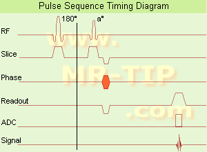
Ultrafast Gradient Echo Sequence Timing Diagram In simple ultrafast GRE imaging, TR and TE are so short, that tissues have a poor imaging signal and - more importantly - poor contrast except when contrast media enhanced (contrast enhanced angiography). Therefore, the magnetization is 'prepared' during the preparation module, most frequently by an initial 180° inversion pulse.
In the pulse sequence timing diagram, the basic ultrafast gradient echo sequence is illustrated. The 180° inversion pulse is executed one time (to the left of the vertical line), the right side represents the data collection period and is often repeated depending on the acquisition parameters.
See also Pulse Sequence Timing Diagram, there you will find a description of the components.
Ultrafast GRE sequences have a short TR,TE, a low flip angle and TR is so short that image acquisition lasts less than 1 second and typically less than 500 ms. Common TR: 3-5 msec, TE: 2 msec, and the flip angle is about 5°. Such sequences are often labeled with the prefix 'Turbo' like TurboFLASH, TurboFFE and TurboGRASS.
This allows one to center the subsequent ultrafast GRE data acquisition around the inversion time TI, where one of the tissues of interest has very little signal as its z-magnetization is passing through zero.
Unlike a standard inversion recovery (IR) sequence, all lines or a substantial segment of k-space image lines are acquired after a single inversion pulse, which can then together be considered as readout module. The readout module may use a variable flip angle approach, or the data acquisition may be divided into multiple segments (shots). The latter is useful particularly in cardiac imaging where acquiring all lines in a single segment may take too long relative to the cardiac cycle to provide adequate temporal resolution.
If multiple lines are acquired after a single pulse, the pulse sequence is a type of gradient echo echo planar imaging (EPI) pulse sequence.

See also Magnetization Prepared Rapid Gradient Echo (MPRAGE) and Turbo Field Echo (TFE).
spacer

• View the DATABASE results for 'Ultrafast Gradient Echo Sequence' (13).Open this link in a new window

MRI Resources 
MRI Accidents - Sequences - Mobile MRI Rental - MRI Training Courses - Image Quality - Intraoperative MRI
 
Incoherent Gradient Echo (RF Spoiled)InfoSheet: - Sequences - 
Intro, 
Overview, 
Types of, 
etc.
 
A gradient echo is generated by using a pair of bipolar gradient pulses. The gradient field is negatively pulsed, causing the spins of the xy-magnetization to dephase. A second gradient pulse is applied with the opposite polarity. During the pulsing, the spins that dephased begin to rephase and generate a gradient echo.
Spoiling can be accomplished by RF or a gradient. The incoherent RF spoiled type of a gradient echo sequence use a continuous shifting of the RF pulse to spoil the residual transverse magnetization. The phase of the RF excitation and receiver channel are varied pseudo randomly with each excitation cycle to prevent the xy magnetization from achieving steady state. T2* does not dominate image contrast, so T1 and PD weighting is practical. This method is effective and can be used to achieve a shorter TR, due to a lack of additional gradients. Spoiling eliminates the effect of the remaining xy-magnetization and leads to steady state longitudinal magnetization. These sequences can be used for breath hold, dynamic imaging and in cine and volume acquisitions.
spacer
Searchterm 'Gradient Echo' was also found in the following services: 
spacer
News  (1)  Resources  (2)  Forum  (6)  
 
Contrast Enhanced Gradient Echo SequenceInfoSheet: - Sequences - 
Intro, 
Overview, 
Types of, 
etc.
 
Contrast enhanced GRE sequences provide T2 contrast but have a relatively poor SNR. Repetitive RF pulses with small flip angles together with appropriate gradient profiles lead to the superposition of two resonance signals.
The first signal is due to the free induction decay FID observed after the first and all ensuing RF excitations.
The second is a resonance signal obtained as a result of a spin echo generated by the second and all addicted RF-pulses.
Hence it is absent after the first excitation, it is a result of the free induction decay of the second to last RF-excitation and has a TE, which is almost 2TR. For this echo to occur the gradients have to be completely symmetrical relative to the half time between two RF-pulses, a condition that makes it difficult to integrate this pulse sequence into a multiple slice imaging technique. The second signal not only contains echo contributions from free induction decay, but obviously weakened by T2-decay. Since the echo is generated by a RF-pulse, it is truly T2 rather than T2* weighted. Correspondingly it is also less sensitive to susceptibility changes and field inhomogeneities.
Companies use different acronyms to describe certain techniques.
Different terms (see also acronyms) for these gradient echo pulse sequences:
CE-FAST Contrast Enhanced Fourier Acquired Steady State,
CE-FFE Contrast Enhanced Fast Field Echo,
CE-GRE Contrast Enhanced Gradient-Echo,
DE-FGR Driven Equilibrium FGR,
FADE FASE Acquisition Double Echo,
PSIF Reverse Fast Imaging with Steady State Precession,
SSFP Steady State Free Precession,
T2 FFE Contrast Enhanced Fast Field Echo (T2 weighted).

In this context, 'contrast enhanced' refers to the pulse sequence, it does not mean enhancement with a contrast agent.
spacer

• View the DATABASE results for 'Contrast Enhanced Gradient Echo Sequence' (4).Open this link in a new window

MRI Resources 
Libraries - - Spine MRI - Musculoskeletal and Joint MRI - IR - Most Wanted
 
Small Tip Angle Gradient Echo T1 WeightedInfoSheet: - Sequences - 
Intro, 
Overview, 
Types of, 
etc.MRI Resource Directory:
 - Sequences -
 
spacer
MRI Resources 
Examinations - Absorption and Emission - Liver Imaging - Hospitals - Functional MRI - Shielding
 
previous      11 - 15 (of 134)     next
Result Pages : [1 2 3 4]  [5 6 7 8 9 10 11 12 13 14 15 16 17 18 19 20 ... ]
 Random Page
 
Share This Page
FacebookTwitterLinkedIn

MR-TIP    
Community   
User
Pass
Forgot your UserID/Password ?    


Personalized protocols (age, gender, body habitus, etc.) lead to :
more automated planning 
improved patient comfort 
better diagnostics 
optimized image quality 
nothing 

Look
      Ups


MR-TIP.com uses cookies! By browsing MR-TIP.com, you agree to our use of cookies.

Magnetic Resonance - Technology Information Portal
Member of SoftWays' Medical Imaging Group - MR-TIP • Radiology-TIP • Medical-Ultrasound-Imaging • 
Copyright © 2003 - 2024 SoftWays. All rights reserved. [ 19 April 2024]
Terms of Use | Privacy Policy | Advertising
 [last update: 2024-02-26 03:41:00]