 | Info
Sheets |
| | | | | | | | | | | | | | | | | | | | | | | | |
 | Out-
side |
| | | | |
|
| | | | | |  | Searchterm 'Gating' was also found in the following services: | | | | |
|  |  |
| |
|

From Hitachi Medical Systems America, Inc.;
the AIRIS made its debut in 1995. Hitachi followed up with the AIRIS II system, which has proven equally successfully. 'All told, Hitachi has installed more than 1,000 MRI systems in the U.S., holding more than 17 percent of the total U.S. MRI installed base, and more than half of the installed base of open MR systems,' says Antonio Garcia, Frost and Sullivan industry research analyst.
Now Altaire employs a blend of innovative Hitachi features called VOSI™ technology, optimizing each sub-system's performance in concert with the
other sub-systems, to give the seamless mix of high-field performance
and the patient comfort, especially for claustrophobic patients, of open MR systems.
Device Information and Specification
CLINICAL APPLICATION
Whole body
DualQuad T/R Body Coil, MA Head, MA C-Spine, MA Shoulder, MA Wrist, MA CTL Spine, MA Knee, MA TMJ, MA Flex Body (3 sizes), Neck, small and large Extremity, PVA (WIP), Breast (WIP), Neurovascular (WIP), Cardiac (WIP) and MA Foot//Ankle (WIP)
SE, GE, GR, IR, FIR, STIR, ss-FSE, FSE, DE-FSE/FIR, FLAIR, ss/ms-EPI, ss/ms EPI- DWI, SSP, MTC, SE/GE-EPI, MRCP, SARGE, RSSG, TRSG, BASG, Angiography: CE, PC, 2D/3D TOF
IMAGING MODES
Single, multislice, volume study
TR
SE: 30 - 10,000msec GE: 3.6 - 10,000msec IR: 50 - 16,700msec FSE: 200 - 16,7000msec
TE
SE : 8 - 250msec IR: 5.2 -7,680msec GE: 1.8 - 2,000 msec FSE: 5.2 - 7,680
0.05 sec/image (256 x 256)
2D: 2 - 100 mm; 3D: 0.5 - 5 mm
Level Range: -2,000 to +4,000
COOLING SYSTEM TYPE
Water-cooled
3.1 m lateral, 3.6 m vertical
| | | | | | | | | | |  Further Reading: Further Reading: | News & More:
|
|
| |
| | | Searchterm 'Gating' was also found in the following services: | | | | |
| | |
| |
|
In the last years, cardiac MRI techniques have progressively improved. No other noninvasive imaging modality provides the same degree of contrast and temporal resolution for the assessment of cardiovascular anatomy and pathology. Contraindications MRI are the same as for other magnetic resonance techniques.
The primary advantage of MRI is extremely high contrast resolution between different tissue types, including blood. Moreover, MRI is a true 3 dimensional imaging modality and images can be obtained in any oblique plane along the true cardiac axes while preserving high temporal and spatial resolution with precise demonstration of cardiac anatomy without the administration of contrast media.
Due to these properties, MRI can precisely characterize cardiac function and quantify cavity volumes, ejection fraction, and left ventricular mass. In addition, cardiac MRI has the ability to quantify flow (see flow quantification), including bulk flow in vessels, pressure gradients across stenosis, regurgitant fractions and shunt fractions. Valve morphology and area can be determined and the severity of stenosis quantified. In certain disease states, such as myocardial infarction, the contrast resolution of MRI is further improved by the addition of extrinsic contrast agents (see myocardial late enhancement).
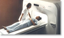
A dedicated cardiac coil, and a field strength higher than 1 Tesla is recommended to have sufficient signal. Cardiac MRI acquires ECG gating. Cardiac gating (ECGs) obtained within the MRI scanner, can be degraded by the superimposed electrical potential of flowing blood in the magnetic field. Therefore, excellent contact between the skin and ECG leads is necessary. For male patients, the skin at the lead sites can be shaved. A good cooperation of the patient is necessary because breath holding at the end of expiration is practiced during the most sequences.
See also Displacement Encoding with Stimulated Echoes.
For Ultrasound Imaging (USI) see Cardiac Ultrasound at Medical-Ultrasound-Imaging.com.
See also the related poll results: ' In 2010 your scanner will probably work with a field strength of' and ' MRI will have replaced 50% of x-ray exams by' | | | | | |
• View the DATABASE results for 'Cardiac MRI' (15).
| | |
• View the NEWS results for 'Cardiac MRI' (15).
| | | | | | Further Reading: | | Basics:
|
|
News & More:
|  |
MRI technology visualizes heart metabolism in real time
Friday, 18 November 2022 by medicalxpress.com | | |
Even early forms of liver disease affect heart health, Cedars-Sinai study finds
Thursday, 8 December 2022 by www.eurekalert.org | | |
MRI sheds light on COVID vaccine-associated heart muscle injury
Tuesday, 15 February 2022 by www.sciencedaily.com | | |
Radiologists must master cardiac CT, MRI to keep pace with demand: The heart is not a magical organ
Monday, 1 March 2021 by www.radiologybusiness.com | | |
Diffusion weighted imaging (DWI) and diffusion tensor imaging (DTI) in the heart (myocardium)
Sunday, 30 August 2020 by github.com | | |
Non-invasive diagnostic procedures for suspected CHD: Search reveals informative evidence
Wednesday, 8 July 2020 by medicalxpress.co | | |
Cardiac MRI Becoming More Widely Available Thanks to AI and Reduced Exam Times
Wednesday, 19 February 2020 by www.dicardiology.com | | |
Controlling patient's breathing makes cardiac MRI more accurate
Friday, 13 May 2016 by www.upi.com | | |
Precise visualization of myocardial injury: World's first patient-based cardiac MRI study using 7T MRI
Wednesday, 10 February 2016 by medicalxpress.com | | |
New technique could allow for safer, more accurate heart scans
Thursday, 10 December 2015 by www.gizmag.com |
|
| |
| | | | | |
| |
|
Cine sequences used in cardiovascular MRI are collection of images (usually at the same spatial location) covering of one full period of cardiac cycle or over several periods in order to obtain complete coverage.
The pulse sequence used, is either a standard gradient echo pulse sequence, a segmented data acquisition, a gradient echo EPI sequence or a gradient echo with balanced gradient waveform.
In cardiac gating studies it is possible to assign consecutive lines either to different images, yielding a multiphase sequence with as many images as lines, or the lines are grouped together into segments and assigned to the same image. The overall time to acquire such a segment has to be small compared to the RR-interval of the cardiac cycle, i. e. 50 ms, and hence contains typically 8 to 16 image lines.
This strategy is called segmented data acquisition, and has the advantage of reducing overall imaging time for cardiac images so that they can be acquired within a breath hold, but obviously decreasing the temporal resolution of each individual image.
This method shows dynamic processes, such as the ejection of blood out of the heart into the aorta, by means of fast imaging and displaying the resulting images in a sequential-loop, the impression of a real-time movie is generated. Ejection fractions and stroke volumes calculated from these cine MRI images in different cardiac axes have been shown to be more accurate than any other imaging modality. See also Cardiac Gating. | | | | | |
• View the DATABASE results for 'Cine Sequence' (2).
| | | | | | Further Reading: | News & More:
|
|
| |
| | | Searchterm 'Gating' was also found in the following services: | | | | |
| | |
| |
|
Device Information and Specification
CLINICAL APPLICATION
Whole body
SE, IR, FSE, FIR, GE, SG, BASG, PBSG, PCIR, DWI, Radial, Angiography: TOF, FLUTE (Fluoro-triggered bolus MRA), Time-resolved MRA
IMAGING MODES
Single, multislice, volume study
Level Range: -2,000 to +4,000
POWER REQUIREMENTS
208/220/240 V, single phase
| | | |
• View the DATABASE results for 'Echelon™ 1.5T' (2).
| | |
• View the NEWS results for 'Echelon™ 1.5T' (3).
| | | | | | Further Reading: | Basics:
|
|
| |
| | | Searchterm 'Gating' was also found in the following services: | | | | |
| | |
| |
|
From Hitachi Medical Systems America, Inc.; because of its dependability, the MRP-7000™ remains popular more than a decade after the first U.S. system was shipped. This system maintains a high resale value, what has made it one of the most sought-after scanners on the used MRI equipment market.
Device Information and Specification CLINICAL APPLICATION Whole body DualQuad T/R Body Coil, MA Head, MA C-Spine, MA Shoulder, MA Wrist, MA CTL Spine, MA Knee, MA TMJ, MA Flex Body (3 sizes), Neck, small and large Extremity, PVA (WIP), Breast (WIP), Neurovascular (WIP), Cardiac (WIP) and MA Foot//Ankle (WIP) SE, GE, GR, IR, FIR, STIR, ss-FSE, FSE, DE-FSE/FIR, FLAIR, ss/ms-EPI, ss/ms EPI- DWI, SSP, MTC, SE/GE-EPI, MRCP, SARGE, RSSG, TRSG, BASG, Angiography: CE, PC, 2D/3D TOFIMAGING MODES Single, multislice, volume study horizontal 2.5 m x 2.1 m vertical | | | |
• View the DATABASE results for 'MRP-7000™' (2).
| | | | |
| | | | |
| | |
| | | |
|
| |
 | Look
Ups |
| |


