Magnetic Resonance - Technology Information Portal Welcome to MRI Technology
Info
  Sheets

Out-
      side


 'Gating' 
SEARCH FOR    
 
  2 3 5 A B C D E F G H I J K L M N O P Q R S T U V W X Y Z
Result : Searchterm 'Gating' found in 4 terms [] and 63 definitions []
previous     46 - 50 (of 67)     next
Result Pages : [1]  [2 3 4 5 6 7 8 9 10 11 12 13 14]
Searchterm 'Gating' was also found in the following services: 
spacer
News  (4)  Resources  (2)  Forum  (1)  
 
MAGNETOM™InfoSheet: - Devices -
Intro, 
Types of Magnets, 
Overview, 
etc.MRI Resource Directory:
 - Devices -
 
www.medical.siemens.com 
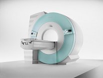
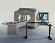
From Siemens Medical Systems;
70 cm + 125 cm + 1.5T and Tim - a combination never seen before in MRI ... MAGNETOM Espree™s unique open bore design can accommodate more types of patients than other 1.5T systems on the market today, in particular the growing population of obese patients. The power of 1.5T combined with Tim technology boosts signal to noise, which is necessary to adequately image obese patients.
Device Information and Specification
CLINICAL APPLICATION
Whole body
CONFIGURATION
Open bore
Body, Tim [32 x 8], Tim [76 coil elements with up to 18 RF channels])
SYNCHRONIZATION
ECG/peripheral: Optional/yes, respiratory gating
PULSE SEQUENCES
GRE, IR, FIR, STIR, TrueIR/FISP, FSE, FLAIR, MT, SS-FSE, MT-SE, MTC, MSE, EPI, 3D DESS//CISS/PSIF, GMR
IMAGING MODES
Single, multislice, volume study, multi angle, multi oblique
SINGLE/MULTI SLICE
Image Processor reconstructing up to 3226 images per second (256 x 256, 25% recFoV)
FOV
35 cm coronal//sagittal, 45 cm axial
Min 2D/3D: 0.1/0.05 mm
1024 x 1024 full screen display
MEASURING MATRIX
64 x 64 to 1024 x 1024
BORE DIAMETER
or W x H
70 cm diameter
MAGNET WEIGHT
3800 kg
H*W*D
? x ? x 125 cm
STRENGTH
33 mT/m
5-GAUSS FRINGE FIELD
2.5 m / 3.8 m
Passive, active
spacer
 
• Related Searches:
    • MRI Equipment
    • Device
    • Hardware
    • Open MRI
    • Superconducting Magnet
 
Further Reading:
  News & More:
First 1.5 Tesla Open Bore MRI Introduced
Tuesday, 10 August 2004   by www.hospimedica.com    
Obesity May Influence Imaging Diagnosis
Wednesday, 22 December 2004   by www.hospimedica.com    
Searchterm 'Gating' was also found in the following services: 
spacer
Radiology  (5) Open this link in a new windowUltrasound  (7) Open this link in a new window
Motion ArtifactInfoSheet: - Artifacts - 
Case Studies, 
Reduction Index, 
etc.MRI Resource Directory:
 - Artifacts -
 
Quick Overview
Please note that there are different common names for this artifact.
Artifact Information
NAME
Motion, phase encoded motion, instability, smearing
DESCRIPTION
Blurring and ghosting
REASON
Movement of the imaged object
HELP
Compensation techniques, more averages, anti spasmodic
Patient motion is the largest physiological effect that causes artifacts, often resulting from involuntary movements (e.g. respiration, cardiac motion and blood flow, eye movements and swallowing) and minor subject movements.
Movement of the object being imaged during the sequence results in inconsistencies in phase and amplitude, which lead to blurring and ghosting. The nature of the artifact depends on the timing of the motion with respect to the acquisition. Causes of motion artifacts can also be mechanical vibrations, cryogen boiling, large iron objects moving in the fringe field (e.g. an elevator), loose connections anywhere, pulse timing variations, as well as sample motion. These artifacts appear in the phase encoding direction, independent of the direction of the motion.
mri safety guidance
Image Guidance
Motion artifacts can be flipped 90° by swapping the phase//frequency encoding directions.
The artifacts can be reduced by using breath holding, cardiac synchronization or respiratory compensation techniques: triggering, gating, retrospective triggering or phase encoding artifact reduction. Flow effects can be reduced by using gradient moment nulling of the first order of flow, gradient moment rephasing or flow compensation, depending of the MRI system.
Peristaltic motion can be reduced with the intravenous injection of an anti-spasmodic (e.g. Buscopan).
By using multiple averages, respiratory motion can be reduced in the same way that multiple averages increase the signal to noise ratio. Noticeable motion averaging is seen when four averages are obtained, six averages are often as good as respiratory compensation techniques and higher averages will continue to improve image quality.
In some cases will help a presaturation of the anatomy that was generating the motion.

See also Phase Encoded Motion Artifact.
spacer

• View the DATABASE results for 'Motion Artifact' (24).Open this link in a new window

 
Further Reading:
  Basics:
The Effects of Breathing Motion on DCE-MRI Images: Phantom Studies Simulating Respiratory Motion to Compare CAIPIRINHA-VIBE, Radial-VIBE, and Conventional VIBE
Tuesday, 7 February 2017   by www.kjronline.org    
  News & More:
Patient movement during MRI: Additional points to ponder
Tuesday, 5 January 2016   by www.healthimaging.com    
Motion-compensation of Cardiac Perfusion MRI using a Statistical Texture Ensemble(.pdf)
June 2003   by www.imm.dtu.dk    
MRI Resources 
MRI Centers - Stimulator pool - Musculoskeletal and Joint MRI - Crystallography - Directories - Mobile MRI Rental
 
OPART™InfoSheet: - Devices -
Intro, 
Types of Magnets, 
Overview, 
etc.MRI Resource Directory:
 - Devices -
 
www3.toshiba.co.jp/medical/products/mr/opart/index.htm From Toshiba America Medical Systems Inc.;
OPART™ is a 0.35 T superconducting open MR system featuring cryogenless operation. Superconducting speed and performance are combined with the patient access advantages of open MRI design for unmatched clinical versatility. OPART™ features innovative technologies such as digital RF, high speed gradients and optimized RF coils, which support advanced MRI applications.
Device Information and Specification
CLINICAL APPLICATION
Whole body
CONFIGURATION
Quadrature, solenoid and multi-channel configurations
SYNCHRONIZATION
ECG/peripheral optional, respiratory gating
PULSE SEQUENCES
SE, FE, IR, FastSE, FastIR, FastFLAIR, Fast STIR, FastFE, FASE, Hybrid EPI, Multi Shot EPI; Angiography: 2D(gate/non-gate)/3D TOF, SORS-STC
IMAGING MODES
Single, multislice, volume study
55 cm, vertical opening
POWER REQUIREMENTS
380/400/415/440/480 V
COOLING SYSTEM TYPE
Cryogenless
STRENGTH
25 mT/m
Passive, active
spacer

• View the DATABASE results for 'OPART™' (2).Open this link in a new window

Searchterm 'Gating' was also found in the following services: 
spacer
News  (4)  Resources  (2)  Forum  (1)  
 
Panorama 0.23T™InfoSheet: - Devices -
Intro, 
Types of Magnets, 
Overview, 
etc.MRI Resource Directory:
 - Devices -
 
www.medical.philips.com/main/products/mri/products/panoramafamily/panorama0.23t_rt/features/ From Philips Medical Systems;
the Panorama 0.23 T, providing a new design optimized for patient comfort, faster reconstruction time than before (300 images/second) and new gradient specifications. Philips' Panorama 0.23 T I/T supports MR-guided interventions, resulting in minimally invasive procedures, more targeted surgery, reduced recovery time and shorter hospital stays. Optional OptoGuide functionality enables real-time needle tracking. Philips' Panorama 0.23 TPanorama 0.2 R/T is the first and only open MRI system to enable radiation therapy planning using MR data sets. The Panorama also features the new and consistent Philips User Interface, an essential element of the Vequion clinical IT family of products and services.
Device Information and Specification
CLINICAL APPLICATION
Whole body
CONFIGURATION
Open MRI/C-arm
Head, head-neck, extremity M-L, neck, body/spine S-XL, shoulder, bilateral breast, wrist, TMJ, flex XS-S-M-L-XL-XXL
SYNCHRONIZATION
ECG/peripheral: Optional/optional, respiratory gating
PULSE SEQUENCES
SE, FE, IR, FFE, DEFFE, DESE, TSE, DETSE, Single shot SE, DRIVE, Balanced FFE, MRCP, Fluid Attenuated Inversion Recovery, Turbo FLAIR, IR-TSE, T1-STIR TSE, T2-STIR TSE, Diffusion Imaging, 3D SE, 3D FFE, MTC;; Angiography: CE-ANGIO, MRA 2D, 3D TOF
IMAGING MODES
Single, multislice, volume study, dynamic, SIMEX, multi chunk 3D, multiple stacks
TR
Min. 6.2 msec
TE
Min. 2.8 msec
SINGLE/MULTI SLICE
50 slices/sec
0.4 cm - 40 cm
1280 X 1024
MEASURING MATRIX
Up to 512 x 512
PIXEL INTENSITY
256 gray scale
MAGNET TYPE
Resistive/iron core
Open x 46 cm x infinite (side-first patient entry)
MAGNET WEIGHT
13110 kg
H*W*D
196 x 121 x 176 cm
POWER REQUIREMENTS
400/480 V
COOLING SYSTEM TYPE
Closed loop chilled water (chiller included)
N/A
STRENGTH
19 mT/m
5-GAUSS FRINGE FIELD
2.4 m / 3.7 m
Passive/active
spacer

• View the DATABASE results for 'Panorama 0.23T™' (2).Open this link in a new window

 
Further Reading:
  News & More:
Magnetic resonance imaging guided musculoskeletal interventions at 0.23T: Chapter 4. Materials and methods
2002
Searchterm 'Gating' was also found in the following services: 
spacer
Radiology  (5) Open this link in a new windowUltrasound  (7) Open this link in a new window
Panorama 0.6TPanorama 0.2InfoSheet: - Devices -
Intro, 
Types of Magnets, 
Overview, 
etc.MRI Resource Directory:
 - Devices -
 
www.medical.philips.com/main/products/mri/products/panoramafamily/panorama0.23t_rt/features/ From Philips Medical Systems;
Panorama 0.6 T is the Philips Mid-Field Open MRI system. It is the most open MR scanner in the market, optimized for patient comfort and faster reconstruction time.
Device Information and Specification
CLINICAL APPLICATION
Whole body
CONFIGURATION
Open MRI/C-arm
Head, head-neck, extremity, neck, body/spine M-XL, shoulder, bilateral breast, wrist, TMJ, flex XS-S-M-L-XL-XXL
SYNCHRONIZATION
ECG/peripheral: Optional/yes, respiratory gating
PULSE SEQUENCES
SE, FE, IR, STIR, FFE, DEFFE, DESE, TSE, DETSE, Single shot SE, DRIVE, Balanced FFE, MRCP, Fluid Attenuated Inversion Recovery, Turbo FLAIR, IR-TSE, T1-STIR TSE, T2-STIR TSE, Diffusion Imaging, 3D SE, 3D FFE, Contrast Perfusion Analysis, MTC;; Angiography: CE-ANGIO, MRA 2D, 3D TOF
IMAGING MODES
Single, multislice, volume study, dynamic, SIMEX, multi chunk 3D, multiple stacks
TR
Min. 4.6 msec
TE
Min. 2.3 msec
SINGLE/MULTI SLICE
50 slices/sec
0.4 cm - 42 cm
1280 X 1024
MEASURING MATRIX
Up to 512 x 512
PIXEL INTENSITY
256 gray scale
MAGNET TYPE
Superconducting / iron core
Open x 47 cm x infinite (side-first patient entry)
MAGNET WEIGHT
38000 kg
H*W*D
254 x 244 x 325 cm
POWER REQUIREMENTS
400/480 V
COOLING SYSTEM TYPE
Liquid helium//air cool
0.00 L/hr helium
STRENGTH
20 mT/m
5-GAUSS FRINGE FIELD
2.4 m / 2.5 m
Passive/active
spacer

• View the DATABASE results for 'Panorama 0.6T™' (2).Open this link in a new window

MRI Resources 
Movies - Calculation - Jobs pool - Nerve Stimulator - Claustrophobia - Knee MRI
 
previous      46 - 50 (of 67)     next
Result Pages : [1]  [2 3 4 5 6 7 8 9 10 11 12 13 14]
 Random Page
 
Share This Page
FacebookTwitterLinkedIn

MR-TIP    
Community   
User
Pass
Forgot your UserID/Password ?    


Personalized protocols (age, gender, body habitus, etc.) lead to :
more automated planning 
improved patient comfort 
better diagnostics 
optimized image quality 
nothing 

Look
      Ups


MR-TIP.com uses cookies! By browsing MR-TIP.com, you agree to our use of cookies.

Magnetic Resonance - Technology Information Portal
Member of SoftWays' Medical Imaging Group - MR-TIP • Radiology-TIP • Medical-Ultrasound-Imaging • 
Copyright © 2003 - 2024 SoftWays. All rights reserved. [ 18 April 2024]
Terms of Use | Privacy Policy | Advertising
 [last update: 2024-02-26 03:41:00]