| Info
Sheets |
| | | | | | | | | | | | | | | | | | | | | | | | |
 | Out-
side |
| | | | |
|
| | | 'Functional Magnetic Resonance Imaging' | |
Result : Searchterm 'Functional Magnetic Resonance Imaging' found in 1 term [ ] and 7 definitions [ ] and 7 definitions [ ], (+ 3 Boolean[ ], (+ 3 Boolean[ ] results ] results
| | previous 6 - 10 (of 11) nextResult Pages : [1] [2] [3] |  | |  | Searchterm 'Functional Magnetic Resonance Imaging' was also found in the following services: | | | | |
| |  |
| |
|
| | | | | |  Further Reading: Further Reading: | | Basics:
|
|
News & More:
| |
| |
| | | Searchterm 'Functional Magnetic Resonance Imaging' was also found in the following services: | | | | |
| | |
| |
|
The ability of magnetic compounds to increase the relaxation
rates of the surrounding water proton spins.
Relaxivity is used to improve the contrast of the image, and
to study tissue specific areas where the contrast agent
better diffuses or to perform functional magnetic resonance imaging.
The relaxivity of MRI contrast agents depends on the
molecular structure and kinetic of the complex.
To increase the number of water molecules that are in the inner sphere
of the complex, or to slow down the molecular rotational
correlation time, are possibilities to improve the water relaxivity.
Relaxivity units ( r1, r2) are mM -1 * sec -1 (at varying temperatures). | | | |
• View the DATABASE results for 'Relaxivity' (51).
| | |
• View the NEWS results for 'Relaxivity' (2).
| | | | | | Further Reading: | News & More:
|
|
| |
| | | | | |
| |
|
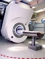
Device Information and Specification
CLINICAL APPLICATION
Whole body
CONFIGURATION
Mobile compact
Whole body, intra-operative head, neck volume, atlas head//neck vascular quadrature phased array, spine quadrature, C/T/L spine phased array, small joint, large joint, TMJ bilateral, shoulder phased array, extremity quadrature volume, wrist, hand quadrature, general purpose flexible, pelvis/abdomen phased array, body quadrature, phased array flexible, breast bilateral
IMAGING MODES
Localizer, single slice, multislice, volume
| | | |
• View the DATABASE results for 'iMotion™ 1.5 Tesla Magnet' (2).
| | | | |
| | | Searchterm 'Functional Magnetic Resonance Imaging' was also found in the following services: | | | | |
| |  |
| |
|
Contrast enhanced MRI is a commonly used procedure in magnetic resonance imaging. The need to more accurately characterize different types of lesions and to detect all malignant lesions is the main reason for the use of intravenous contrast agents.
Some methods are available to improve the contrast of different tissues. The focus of dynamic contrast enhanced MRI (DCE-MRI) is on contrast kinetics with demands for spatial resolution dependent on the application. DCE- MR imaging is used for diagnosis of cancer (see also liver imaging, abdominal imaging, breast MRI, dynamic scanning) as well as for diagnosis of cardiac infarction (see perfusion imaging, cardiac MRI). Quantitative DCE-MRI requires special data acquisition techniques and analysis software.
Contrast enhanced magnetic resonance angiography (CE-MRA) allows the visualization of vessels and the temporal resolution provides a separation of arteries and veins. These methods share the need for acquisition methods with high temporal and spatial resolution.
Double contrast administration (combined contrast enhanced (CCE) MRI) uses two contrast agents with complementary mechanisms e.g., superparamagnetic iron oxide to darken the background liver and gadolinium to brighten the vessels. A variety of different categories of contrast agents are currently available for clinical use.
Reasons for the use of contrast agents in MRI scans are:
•
Relaxation characteristics of normal and pathologic tissues are not always different enough to produce obvious differences in signal intensity.
•
Pathology that is sometimes occult on unenhanced images becomes obvious in the presence of contrast.
•
Enhancement significantly increases MRI sensitivity.
•
In addition to improving delineation between normal and abnormal tissues, the pattern of contrast enhancement can improve diagnostic specificity by facilitating characterization of the lesion(s) in question.
•
Contrast can yield physiologic and functional information in addition to lesion delineation.
Common Indications:
Brain MRI : Preoperative/pretreatment evaluation and postoperative evaluation of brain tumor therapy, CNS infections, noninfectious inflammatory disease and meningeal disease.
Spine MRI : Infection/inflammatory disease, primary tumors, drop metastases, initial evaluation of syrinx, postoperative evaluation of the lumbar spine: disk vs. scar.
Breast MRI : Detection of breast cancer in case of dense breasts, implants, malignant lymph nodes, or scarring after treatment for breast cancer, diagnosis of a suspicious breast lesion in order to avoid biopsy.
For Ultrasound Imaging (USI) see Contrast Enhanced Ultrasound at Medical-Ultrasound-Imaging.com.
See also Blood Pool Agents, Myocardial Late Enhancement, Cardiovascular Imaging, Contrast Enhanced MR Venography, Contrast Resolution, Dynamic Scanning, Lung Imaging, Hepatobiliary Contrast Agents, Contrast Medium and MRI Guided Biopsy. | | | | | | | | | | |
• View the DATABASE results for 'Contrast Enhanced MRI' (14).
| | |
• View the NEWS results for 'Contrast Enhanced MRI' (8).
| | | | | | Further Reading: | Basics:
|
|
News & More:
|  |
FDA Approves Gadopiclenol for Contrast-Enhanced Magnetic Resonance Imaging
Tuesday, 27 September 2022 by www.pharmacytimes.com | | |
Effect of gadolinium-based contrast agent on breast diffusion-tensor imaging
Thursday, 6 August 2020 by www.eurekalert.org | | |
Artificial Intelligence Processes Provide Solutions to Gadolinium Retention Concerns
Thursday, 30 January 2020 by www.itnonline.com | | |
Accuracy of Unenhanced MRI in the Detection of New Brain Lesions in Multiple Sclerosis
Tuesday, 12 March 2019 by pubs.rsna.org | | |
The Effects of Breathing Motion on DCE-MRI Images: Phantom Studies Simulating Respiratory Motion to Compare CAIPIRINHA-VIBE, Radial-VIBE, and Conventional VIBE
Tuesday, 7 February 2017 by www.kjronline.org | | |
Novel Imaging Technique Improves Prostate Cancer Detection
Tuesday, 6 January 2015 by health.ucsd.edu | | |
New oxygen-enhanced MRI scan 'helps identify most dangerous tumours'
Thursday, 10 December 2015 by www.dailymail.co.uk | | |
All-organic MRI Contrast Agent Tested In Mice
Monday, 24 September 2012 by cen.acs.org | | |
A groundbreaking new graphene-based MRI contrast agent
Friday, 8 June 2012 by www.nanowerk.com |
|
| |
| | | Searchterm 'Functional Magnetic Resonance Imaging' was also found in the following services: | | | | |
| | |
| |
|
Special imaging primarily means advanced MRI techniques used for qualitative and quantitative measurement of biological metabolism as e.g., spectroscopy, perfusion imaging (PWI, ASL), diffusion weighted imaging ( DWI, DTI, DTT) and brain function ( BOLD, fMRI). This physiological magnetic resonance techniques offer insights into brain structure, function, and metabolism.
Spectroscopy provides functional information related to identification and quantification of e.g. brain metabolites.
MR perfusion imaging has applications in stroke, trauma, and brain neoplasm. MRI provides the high spatial and temporal resolution needed to measure blood flow to the brain. arterial spin labeling techniques utilize the intrinsic protons of blood and brain tissue, labeled by special preparation pulses, rather than exogenous tracers injected into the blood.
MR diffusion tensor imaging characterizes the ability of water to spread across the brain in different directions. Diffusion parallel to nerve fibers has been shown to be greater than diffusion in the perpendicular direction. This provides a tool to study in vivo fiber connectivity in brain MRI.
FMRI allows the detection of a functional activation in the brain because cortical activity is intimately related to local metabolism changes. See also Diffusion Tensor Tractography. | | | |
• View the NEWS results for 'Special Imaging' (14).
| | | | | | Further Reading: | Basics:
|
|
News & More:
| |
| |
| | | | |
| | | |
|
| |
 | Look
Ups |
| |