Magnetic Resonance - Technology Information Portal Welcome to MRI Technology
Info
  Sheets

Out-
      side


 'Field Strength' 
SEARCH FOR    
 
  2 3 5 A B C D E F G H I J K L M N O P Q R S T U V W X Y Z
Result : Searchterm 'Field Strength' found in 1 term [] and 135 definitions []
previous     11 - 15 (of 136)     next
Result Pages : [1]  [2 3 4 5 6 7 8 9 10 11 12 13 14 15 16 17 18 19 20 ... ]
Searchterm 'Field Strength' was also found in the following services: 
spacer
News  (13)  Resources  (3)  Forum  (7)  
 
Chemical Shift ArtifactInfoSheet: - Artifacts - 
Case Studies, 
Reduction Index, 
etc.MRI Resource Directory:
 - Artifacts -
 
Quick Overview
Please note that there are different common names for this artifact.
Artifact Information
NAME
Chemical shift, black boundary, spatial misregistration, relief
DESCRIPTION
Black or bright band
During frequency encoding, fat protons precess slower than water protons in the same slice because of their magnetic shielding. Through the difference in resonance frequency between water and fat, protons at the same location are misregistrated (dislocated) by the Fourier transformation, when converting MRI signals from frequency to spatial domain. This chemical shift misregistration cause accentuation of any fat-water interfaces along the frequency axis and may be mistaken for pathology. Where fat and water are in the same location, this artifact can be seen as a bright or dark band at the edge of the anatomy.
Protons in fat and water molecules are separated by a chemical shift of about 3.5 ppm. The actual shift in Hertz (Hz) depends on the magnetic field strength of the magnet being used. Higher field strength increases the misregistration, while in contrast a higher gradient strength has a positive effect. For a 0.3 T system operating at 12.8 MHz the shift will be 44.8 Hz compared with a 223.6 Hz shift for a 1.5 T system operating at 63.9 MHz.
mri safety guidance
Image Guidance
For artifact reduction helps a smaller water fat shift (higher bandwidth), a higher matrix, an in phase TE or a spin echo technique. Since the misregistration offset is present in the read out axis the patient may be rescanned with this axis parallel to the fat-water interface. Steeper gradient may be employed to reduce the chemical shift offset in mm. Another strategy is to employ specialized pulse sequences such as fat saturation or inversion recovery imaging. Fat suppression techniques eliminate chemical shift artifacts caused by the lack of fat signal.

See also Black Boundary Artifact and Magnetic Resonance Spectroscopy.
spacer
 
• Related Searches:
    • Chemical Shift Imaging
    • Spatial Misregistration Artifact
    • Dual Echo Fast Gradient Echo
    • Dual Echo Sequence
    • Chemical Shift
 
Further Reading:
  Basics:
MRI Artifact Gallery
   by chickscope.beckman.uiuc.edu    
  News & More:
What is chemical shift artefact? Why does it occur? How many Hz at 1.5 T?
   by www.revisemri.com    
Abdominal MRI at 3.0 T: The Basics Revisited
Wednesday, 20 July 2005   by www.ajronline.org    
Searchterm 'Field Strength' was also found in the following services: 
spacer
Radiology  (5) Open this link in a new windowUltrasound  (2) Open this link in a new window
Fringe Field
 
The magnetic field outside the magnet, which is also called the stray field. Its strength depends on the magnet type and the field strength. The higher the field strength, the larger the fringe field.

See Magnetic Fringe Field.
spacer

• View the DATABASE results for 'Fringe Field' (57).Open this link in a new window

 
Further Reading:
  Basics:
Physics of MRI Safety
   by www.aapm.org    
MRI Resources 
Health - Jobs - Journals - General - Claustrophobia - Most Wanted
 
High Field MRI
 
The principal advantage of MRI at high field is the increase in signal to noise ratio. This can be used to improve anatomic and/or temporal resolution and reduce scan time while preserving image quality. MRI devices for whole body imaging for human use are available up to 3 tesla (3T). Functional MRI (fMRI) and MR spectroscopy (MRS) benefit significantly. In addition, 3T machines have a great utility in applications such as TOF MRA and DTI. Higher field strengths are used for imaging of small parts of the body or scientific animal experiments. Higher contrast may permit reduction of gadolinium doses and, in some cases, earlier detection of disease.
Using high field MRI//MRS, the RF-wavelength and the dimension of the human body complicating the development of MR coils. The absorption of RF power causes heating of the tissue. The energy deposited in the patient's tissues is fourfold higher at 3T than at 1.5T. The specific absorption rate (SAR) induced temperature changes of the human body are the most important safety issue of high field MRI//MRS.
Susceptibility and chemical shift dispersion increase like T1, therefore high field MRI occasionally exhibits imaging artifacts. Most are obvious and easily recognized but some are subtle and mimic diseases. A thorough understanding of these artifacts is important to avoid potential pitfalls. Some imaging techniques or procedures can be utilized to remove or identify artifacts.

See also Diffusion Tensor Imaging.

See also the related poll result: 'In 2010 your scanner will probably work with a field strength of'
Medical-Ultrasound-Imaging.comMagnetic Resonance Guided Focused Ultrasound,  High Intensity Focused Ultrasound
spacer

• View the DATABASE results for 'High Field MRI' (16).Open this link in a new window


• View the NEWS results for 'High Field MRI' (9).Open this link in a new window.
 
Further Reading:
  Basics:
Next-generation 7 T scanner ramps the resolution of brain MR imaging
Wednesday, 17 January 2024   by physicsworld.com    
A paired dataset of T1- and T2-weighted MRI at 3 Tesla and 7 Tesla
Thursday, 27 July 2023   by www.nature.com    
CLINICAL WHOLE BODY MRI AT 3.0 T(.pdf)
2001
Musculoskeletal MRI at 3.0 T: Relaxation Times and Image Contrast
Sunday, 1 August 2004   by www.ajronline.org    
  News & More:
How safe is 7T MRI for patients with neurosurgical implants?
Thursday, 17 November 2022   by healthimaging.com    
Impact of Magnetic Field Inhomogeneity on the Quality of Magnetic Resonance Images and Compensation Techniques: A Review
Saturday, 1 October 2022   by www.dovepress.com    
7-T clinical MRI of the shoulder in patients with suspected lesions of the rotator cuff
Friday, 7 February 2020   by eurradiolexp.springeropen.com    
A 100-hour MRI scan captured the most detailed look yet at a whole human brain
Monday, 8 July 2019   by www.sciencenews.or    
T2-Weighted Liver MRI Using the MultiVane Technique at 3T: Comparison with Conventional T2-Weighted MRI
Friday, 16 October 2015   by www.ncbi.nlm.nih.gov    
Ultra-high-field MRI reveals language centres in the brain in much more detail
Tuesday, 28 October 2014   by medicalxpress.com    
Turbo-FLASH Based Arterial Spin Labeled Perfusion MRI at 7 T
Thursday, 20 June 2013   by www.plosone.org    
High-Resolution, Spin-Echo BOLD, and CBF fMRI at 4 and 7 T(.pdf)
October 2002   by otg.downstate.edu    
Vascular Filters of Functional MRI: Spatial Localization Using BOLD and CBV Contrast
Searchterm 'Field Strength' was also found in the following services: 
spacer
News  (13)  Resources  (3)  Forum  (7)  
 
Low Field MRIForum -
related threads
 
Advantages of low field imaging are the small-sized 5 Gauss fringe field and therefore the less static magnetic field exposure for the surrounding area, as well as less contraindications causing lower risks for the MRI safety by implemented metal and magnetic devices and equipment.
Low field systems are sometimes for restricted use, e.g. dedicated extremity scanner or open MRI devices. Open MRI devices equipped with permanent magnets are well-suited for MR guided interventions because these machines combine the lower magnetic fields of this type of magnets and the better patient access of open MRI scanner.
In some cases, the contrast of different tissues is better at lower field strength, depending on their T1 or T2 relaxation times. The disadvantage of the lower signal to noise ratio are a poor resolution and a longer scan time for a good image quality.

See also Claustrophobia, Contraindications and MRI Safety.

See also the related poll result: 'In 2010 your scanner will probably work with a field strength of'
spacer

• View the DATABASE results for 'Low Field MRI' (8).Open this link in a new window


• View the NEWS results for 'Low Field MRI' (5).Open this link in a new window.
 
Further Reading:
  Basics:
Improving portable low-field MRI image quality through image-to-image translation using paired low- and high-field images
Friday, 1 December 2023   by www.nature.com    
  News & More:
Safety of Bedside Portable Low-Field Brain MRI in ECMO Patients Supported on Intra-Aortic Balloon Pump
Friday, 18 November 2022   by www.mdpi.com    
Researchers at the University of Tsukuba develop a portable MRI system specifically for identifying wrist cartilage damage among athletes, providing a convenient means of early detection and treatment of injuries
Tuesday, 26 April 2022   by www.tsukuba.ac.jp    
This bizarre looking helmet can create better brain scans
Friday, 11 February 2022   by www.sciencedaily.com    
A low-cost and shielding-free ultra-low-field brain MRI scanner
Tuesday, 14 December 2021   by www.nature.com    
Portable MRI provides life-saving information to doctors treating strokes
Thursday, 5 August 2021   by news.yale.edu    
Synaptive Evry, an MRI for Any Space, Cleared by FDA
Thursday, 30 April 2020   by www.medgadget.com    
World's First Portable MRI Cleared by FDA
Monday, 17 February 2020   by www.medgadget.com    
Introducing a point-of-care MRI system
Tuesday, 29 October 2019   by healthcare-in-europe.com    
Opportunities in Interventional and Diagnostic Imaging by Using High-performance Low-Field-Strength MRI
Tuesday, 1 October 2019   by pubs.rsna.org    
Portable 'battlefield MRI' comes out of the lab
Thursday, 30 April 2015   by physicsworld.com    
Portable MRI could aid wounded soldiers and children in the third world
Thursday, 23 April 2015   by phys.org    
Searchterm 'Field Strength' was also found in the following services: 
spacer
Radiology  (5) Open this link in a new windowUltrasound  (2) Open this link in a new window
MAGNETOM Harmonyâ„¢InfoSheet: - Devices -
Intro, 
Types of Magnets, 
Overview, 
etc.MRI Resource Directory:
 - Devices -
 
www.med.siemens.com/med/d/gg/mr/products/harmony.htm From Siemens Medical Systems;
the MAGNETOM Harmony system is a high-performance MR imaging system based on 1.0 T field strength for best clinical routine MRI. It combines patient-friendly design features with the benefits of short imaging time, high anatomical resolution, and highest productivity.
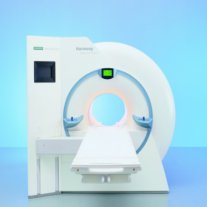
Device Information and Specification
CLINICAL APPLICATION
Whole body
CONFIGURATION
Compact
Body, head, spine, knee, neck, TMJ, extremity, breast, shoulder, others
Yes/SVS opt.
SYNCHRONIZATION
ECG/peripheral, respiratory gating
PULSE SEQUENCES
GRE, IR, FIR, STIR, TrueIR/FISP, FSE, FLAIR, MT, SS-FSE, MT-SE, MTC, MSE, EPI, GMR, fat/water sat./exc.
IMAGING MODES
Single, multislice, volume study, multi angle, multi oblique
TR
2.4 msec std.; 2.0 opt.; 1.8 w/ 30mT/m at 256matrix
TE
1.1 msec std.; 0.9 opt.; 0.78 w/30 mT/m at 256matrix
SINGLE/MULTI SLICE
178 images/sec at 256 x 256 at 100% FOV
FOV
0.5 cm - 50 cm
Min 2D/3D: 0.1/0.05 mm
1024 x 1024 full screen display
MEASURING MATRIX
64 x 64 to 1024 x 1024
21 micrometer in plane, 11 micrometer optional
BORE DIAMETER
or W x H
60 cm
MAGNET WEIGHT (gantry included)
3500kg, 5000kg in operation
DIMENSION H*W*D (gantry included)
POWER REQUIREMENTS
380/400/420/440/480 V
COOLING SYSTEM TYPE
Single cryogen, 2 stage refrig.
CRYOGEN USE
Liquid He
STRENGTH
20/35 mT/m standard, 30/52 opt.
5-GAUSS FRINGE FIELD
2.5 m / 3.9 m
Passive, act.; 1st order std./2nd opt.
spacer

• View the DATABASE results for 'MAGNETOM Harmony™' (2).Open this link in a new window

 
Further Reading:
  Basics:
Harmony Brochure(.pdf)
MRI Resources 
Examinations - MRCP - MRI Centers - RIS - MRA - Pregnancy
 
previous      11 - 15 (of 136)     next
Result Pages : [1]  [2 3 4 5 6 7 8 9 10 11 12 13 14 15 16 17 18 19 20 ... ]
 Random Page
 
Share This Page
FacebookTwitterLinkedIn

MR-TIP    
Community   
User
Pass
Forgot your UserID/Password ?    


New acceleration techniques will :
reduce scan times 
cause artifacts 
increase expenses 
be useful if you have a lot of experience 
doesn't do much 
never heard of 

Look
      Ups


MR-TIP.com uses cookies! By browsing MR-TIP.com, you agree to our use of cookies.

Magnetic Resonance - Technology Information Portal
Member of SoftWays' Medical Imaging Group - MR-TIP • Radiology-TIP • Medical-Ultrasound-Imaging • 
Copyright © 2003 - 2024 SoftWays. All rights reserved. [ 23 April 2024]
Terms of Use | Privacy Policy | Advertising
 [last update: 2024-02-26 03:41:00]