Magnetic Resonance - Technology Information Portal Welcome to MRI Technology
Info
  Sheets

Out-
      side


 'Field Strength' 
SEARCH FOR    
 
  2 3 5 A B C D E F G H I J K L M N O P Q R S T U V W X Y Z
Result : Searchterm 'Field Strength' found in 1 term [] and 135 definitions []
previous     56 - 60 (of 136)     next
Result Pages : [1]  [2 3 4 5 6 7 8 9 10 11 12 13 14 15 16 17 18 19 20 ... ]
Searchterm 'Field Strength' was also found in the following services: 
spacer
News  (13)  Resources  (3)  Forum  (7)  
 
Image Quality
 
The quality of magnetic resonance imaging is particularly dependent on image resolution (matrix, field of view, slice thickness), contrast (TE, TR), signal to noise ratio (bandwidth, signal averaging) and lack of artifacts. These MRI parameters are affected by the field homogeneity, the field strength, the coil, the pulse sequence type and imaging techniques like parallel imaging.

See also Image Contrast Characteristics.
spacer
 
• Related Searches:
    • Signal to Noise Ratio
    • Resolution
    • Lung Imaging
    • Cervical Spine MRI
    • Low Field MRI
 
Further Reading:
  Basics:
Image Characteristics and Quality
   by www.sprawls.org    
  News & More:
Impact of Magnetic Field Inhomogeneity on the Quality of Magnetic Resonance Images and Compensation Techniques: A Review
Saturday, 1 October 2022   by www.dovepress.com    
Researchers design 'intelligent' metamaterial to make MRIs affordable and accessible
Tuesday, 5 November 2019   by phys.org    
Patient movement during MRI: Additional points to ponder
Tuesday, 5 January 2016   by www.healthimaging.com    
Novel Imaging Technique Improves Prostate Cancer Detection
Tuesday, 6 January 2015   by health.ucsd.edu    
Searchterm 'Field Strength' was also found in the following services: 
spacer
Radiology  (5) Open this link in a new windowUltrasound  (2) Open this link in a new window
In Phase Image
 
The term in phase refers to an image in which the signals from two spectral components (such as fat and water) add constructively in a voxel. T1 weighted in phase images are acquired by a gradient echo-based technique with a short TR, TE and a high flip angle greater than 60 degrees. To some degree, in phase sequences are more sensitive to detection of focal hepatic lesions than out of phase for evaluating reduced lesion-to-liver contrast, but the choice for a T1 gradient echo sequence is still based on field strength, advanced imaging techniques (breath hold imaging), and physician preference.
 
Images, Movies, Sliders:
 MRI Liver In Phase  Open this link in a new window
    
 
spacer

• View the DATABASE results for 'In Phase Image' (4).Open this link in a new window

 
Further Reading:
  News & More:
Direct Water and Fat Determination in Two-Point Dixon Imaging
April 2013   by scholarship.rice.edu    
MRI Resources 
MRI Technician and Technologist Jobs - Implant and Prosthesis - Shoulder MRI - Safety Products - Diffusion Weighted Imaging - Pregnancy
 
Incoherent Gradient Echo (Gradient Spoiled)InfoSheet: - Sequences - 
Intro, 
Overview, 
Types of, 
etc.
 
The incoherent gradient echo (gradient spoiled) type of sequence uses a continuous shifting of the RF pulse to spoil the remaining transverse magnetization. The transverse magnetization is destroyed by a magnetic field gradient. This results in a T1 weighted image. Spoiling can be accomplished by RF or a gradient.
Gradient spoiling occurs after each echo by using strong gradients in the slice-select direction after the frequency encoding and before the next RF pulse. Because spins in different locations in the magnet thereby experience a variety of magnetic field strengths, they will precess at differing frequencies; as a consequence they will quickly become dephased. Magnetic field gradients are not very efficient at spoiling the transverse steady state. To be effective, the spins must be forced to precess far enough to become phased randomly with respect to the RF excitation pulse. In clinical MRI machines, the field gradients are set up in such a way that they increase and decrease relative to the center of the magnet; the magnetic field at the magnet 'isocenter' does not change.
The T1 weighting increases with the flip angle and the T2* weighting increases with echo time (TE). Typical repetition time (TR) are 30-500 ms and TE less than 15 ms.

See also Ernst Angle.
spacer
Searchterm 'Field Strength' was also found in the following services: 
spacer
News  (13)  Resources  (3)  Forum  (7)  
 
Infinion 1.5TPanorama 0.2InfoSheet: - Devices -
Intro, 
Types of Magnets, 
Overview, 
etc.MRI Resource Directory:
 - Devices -
 
From Philips Medical Systems; www.medical.philips.com/main/products/mri/products/infinion1.5t/features/ 
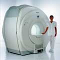
Philips Infinion 1.5 T is designed to maximize the efficiency and quality of patient care. Developed with the patient in mind, the Infinion is the shortest and most open 1.5T scanner available. The unique 'ultra short' 1.4 m magnet assures patient comfort and acceptance without compromising image quality and clinical performance.
Device Information and Specification
CLINICAL APPLICATION
Whole body
CONFIGURATION
Ultra short bore
Head, head / neck, integrated C-spine, L/T spine array, small large GP coils, body flex array, torso pelvis array, breast array, endocavitary, shoulder array, lower extremity, hand / wrist, cardiac, PV array
SYNCHRONIZATION
ECG/peripheral, respiratory gating
PULSE SEQUENCES
SE, TSE, SS TSE, EPI, IR, STIR, FLAIR, FFE, TFE, T1 TFE, T2 TFE, Presat, Fatsat, MTC, Diff-opt., Angiography: PCA, MCA, TOF
IMAGING MODES
Single slice, single volume, multi slice, multi volume
TR
3.1 msec minimum
TE
0.9 msec minimum
SINGLE/MULTI SLICE
80 images/sec std.; up to320 opt.@256
FOV
0.4 - 56 cm
2D: 0.3 mm; 3D: 0.2 mm
1024 x 1024
MEASURING MATRIX
8 x 8 to 1024 x 1024
PIXEL INTENSITY
256 gray scale
BORE DIAMETER
or W x H
60 cm diameter (patient)
MAGNET WEIGHT
4100 kg w/cryogen's
H*W*D
233 (lead fitted) x 198 x 140 cm
POWER REQUIREMENTS
400/480 V
COOLING SYSTEM TYPE
Closed loop, chilled water
CRYOGEN USE
0.06 L/hr helium
STRENGTH
30 mT/m
5-GAUSS FRINGE FIELD
3.0 m / 5.0 m
Passive/active
spacer

• View the DATABASE results for 'Infinion 1.5T™' (2).Open this link in a new window

Searchterm 'Field Strength' was also found in the following services: 
spacer
Radiology  (5) Open this link in a new windowUltrasound  (2) Open this link in a new window
Intera 0.5TPanorama 0.2InfoSheet: - Devices -
Intro, 
Types of Magnets, 
Overview, 
etc.MRI Resource Directory:
 - Devices -
 
www.medical.philips.com/de/products/mri/products/ From Philips Medical Systems;
this low field MRI system of the Intera-family is onto diagnostic safety and flexibility aligned - cost effective and with high patient acceptance.
Device Information and Specification
CLINICAL APPLICATION
Whole body
CONFIGURATION
Short bore compact
Standard: head, body, C1, C3; Optional: Small joint, flex-E, flex-R, endocavitary (L and S), dual TMJ, knee, neck, T/L spine, breast; Optional phased array: Spine, pediatric, 3rd party connector, Flex-S-M-L, flex body, flex cardiac, neuro-vascular, head
SYNCHRONIZATION
ECG/peripheral: Optional/yes, respiratory gating
PULSE SEQUENCES
SE, Modified-SE (TSE), DAVE, STIR, FLAIR, SPIR, MTC, Dynamic, Keyhole, CLEAR, Q Flow, Balanced FFE, Multi Chunk 3D, Multi Stack 3D, FFE-EPI, SE-EPI, IR-EPI, GRASE, Diffusion Imaging, Perfusion Imaging;; Angiography: Inflow MRA, TONE, PCA, CE MRA
IMAGING MODES
Single Slice 2D , Multi Single Slice 2D, Multi Slice 2D, 3D, Multi Chunk 3D, Multi Stack 3D
TR
Min. 2.9 (Omni) msec
TE
Min. 1.0 (Omni) msec
SINGLE/MULTI SLICE
RapidView Recon. greater than 500 @ 256 Matrix
FOV
Max. 53 cm
0.1 mm (Omni)
128 x 128, 256 x 256,512 x 512,1024 x 1024
MEASURING MATRIX
Variable in 1% increments
PIXEL INTENSITY
Lum.: 120 cd/m2; contrast: 150:1
Variable (op. param. depend.)
60 x 60 cm
MAGNET WEIGHT
2500 kg
H*W*D
240 x 188 x 157 cm
POWER REQUIREMENTS
380/400 V
CRYOGEN USE
0.03 L/hr helium
STRENGTH
23 mT/m (Omni)
5-GAUSS FRINGE FIELD
2.1 m / 2.8 m
Passive and dynamic
spacer

• View the DATABASE results for 'Intera 0.5T™' (2).Open this link in a new window

MRI Resources 
MR Myelography - Coils - - Research Labs - Safety Products - Education pool
 
previous      56 - 60 (of 136)     next
Result Pages : [1]  [2 3 4 5 6 7 8 9 10 11 12 13 14 15 16 17 18 19 20 ... ]
 Random Page
 
Share This Page
FacebookTwitterLinkedIn

MR-TIP    
Community   
User
Pass
Forgot your UserID/Password ?    


Personalized protocols (age, gender, body habitus, etc.) lead to :
more automated planning 
improved patient comfort 
better diagnostics 
optimized image quality 
nothing 

Look
      Ups


MR-TIP.com uses cookies! By browsing MR-TIP.com, you agree to our use of cookies.

Magnetic Resonance - Technology Information Portal
Member of SoftWays' Medical Imaging Group - MR-TIP • Radiology-TIP • Medical-Ultrasound-Imaging • 
Copyright © 2003 - 2024 SoftWays. All rights reserved. [ 20 April 2024]
Terms of Use | Privacy Policy | Advertising
 [last update: 2024-02-26 03:41:00]