Magnetic Resonance - Technology Information Portal Welcome to MRI Technology
Info
  Sheets

Out-
      side


 'Device' 
SEARCH FOR    
 
  2 3 5 A B C D E F G H I J K L M N O P Q R S T U V W X Y Z
Result : Searchterm 'Device' found in 4 terms [] and 130 definitions []
previous     16 - 20 (of 134)     next
Result Pages : [1]  [2 3 4 5 6 7 8 9 10 11 12 13 14 15 16 17 18 19 20 ... ]
Searchterm 'Device' was also found in the following services: 
spacer
News  (96)  Resources  (22)  Forum  (10)  
 
GE LunarMRI Resource Directory:
 - Manufacturers -
 
www.gehealthcare.com [This entry is marked for removal.]

General Electric (GE) agreed to buy diagnostic systems maker Lunar Corp. for $150m. in March 2000. In 2004/05 it seems that the integration process into GE Healthcare has been completed. (GE Medical Systems and Amersham announced in April 2004 the completion of a share exchange acquisition of Amersham Health by GE. The result of this acquisition is the new GE Healthcare, based in the UK, totally owned by General Electric (GE).

The U.S.-based company developed bone densitometers and scanning machines that measure bone density as a way of diagnosing osteoporosis and other metabolic bone diseases. GE Lunar marketed these products worldwide.
GE Lunar announced a distribution agreement with MagneVu for domestic sales of the MagneVu 1000, a portable MRI device for orthopedic use, under the trade name Applause™.
GE Lunar was the exclusive U.S. distributor of MR-devices manufactured by Esaote S.p.A. These compact in-office MRI™ machines are designed to fit all practice sizes in orthopedic imaging and complete the range of diagnostic imaging systems.
spacer
 
Further Reading:
  News & More:
Portable MR system aids diagnosis of rheumatic diseases
Sunday, 12 January 2003   by www.diagnosticimaging.com    
Searchterm 'Device' was also found in the following services: 
spacer
Radiology  (21) Open this link in a new windowUltrasound  (74) Open this link in a new window
Hardware
 
MRI hardware includes the electrical and mechanical components of a scanning device.
The main hardware components for the MRI machine are:
The magnet establishing the B0 field to align the spins.
Within the magnet are the gradient coils for producing variations in B0 in the X, Y, and Z directions to make a localization of the received data possible.
Within the gradient coil or directly on the object being imaged is the radio frequency (RF) coil. This RF coil is used to establish the B1 magnetic field necessary to excite the spinning nuclei. The RF coil also detects the signal emitted from the spins within the object being imaged.
The RF amplifier increases the power of the pulses.
The analog to digital converter converts the received analog raw data into digital values.
Depending on the design of the device and the body part being imaged the patient is positioned inside the magnet (e.g. on a movable table or standing upright).
The MRI scan room is surrounded by a RF shield (Faraday cage).
In addition, a computer console, a display, and a film printer belong to the MRI equipment.

See also the related poll result: 'Most outages of your scanning system are caused by failure of'
Radiology-tip.comradCT Scanner,  Gamma Camera
spacer
Medical-Ultrasound-Imaging.comUltrasound Machine
spacer

• View the DATABASE results for 'Hardware' (19).Open this link in a new window


• View the NEWS results for 'Hardware' (1).Open this link in a new window.
 
Further Reading:
  Basics:
Imaging Hardware
   by www.fmrib.ox.ac.uk    
  News & More:
Why non-magnetic capacitors matter in medical imaging
Wednesday, 19 February 2020   by www.medicaldesignandoutsourcing.com    
A transportable MRI machine to speed up the diagnosis and treatment of stroke patients
Wednesday, 22 April 2015   by medicalxpress.com    
Magnetic resonance angiography: current status and future directions
Wednesday, 9 March 2011   by www.jcmr-online.com    
MRI Resources 
MRI Technician and Technologist Jobs - Liver Imaging - PACS - MRI Reimbursement - MR Myelography - Claustrophobia
 
MAGNETOM 7T
 
www.healthcare.siemens.com/magnetic-resonance-imaging/7t-mri-scanner/magnetom-7t 
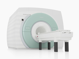
From Siemens Medical Systems;
The MAGNETOM 7T is designed as an open research platform. 7T MRI provides anatomical detail at the submillimiter scale, enhanced contrast mechanisms, outstanding spectroscopy performance, ultra-high resolution functional imaging (fMRI), multinuclear whole-body MRI and functional information.
This ultra high field (UHF) MRI device is a research system and not cleared, approved or licensed in any jurisdiction for patient examinations.
Device Information and Specification
CLINICAL APPLICATION
Whole body
CONFIGURATION
Compact
7 Tesla
High-performance, ultra high field coils available. Integration and support for coil developments.
CHANNELS (min. / max. configuration)
32, optional 8 channels TX array
Chemical shift imaging, single voxel spectroscopy, multinuclear imaging optional
IMAGING TECHNIQUES
iPAT, mSENSE and GRAPPA (image, k-space), noncontrast angiography, plaque imaging, radial motion compensation
FOV
40 x 40 x 30 cm³ less than 8% nonlinearity
BORE DIAMETER
or W x H
60 cm
TABLE CAPACITY
200 kg
MAGNET WEIGHT (gantry included)
35017 kg
DIMENSION H*W*D (gantry included)
320 x 240 x 317,5 cm
5-GAUSS FRINGE FIELD
7.9 m / 5.6 m
CRYOGEN USE
Zero boil off rate
COOLING SYSTEM
Water
up to 200 T/m/s
MAX. AMPLITUDE
up to 70 mT/m
Up to 3rd order shim coils, user configurable B0 shim ? B0 maps and ROI definition
POWER REQUIREMENTS
2000 Volts, 650A
spacer
 
Further Reading:
  Basics:
MAGNETOM 7T Product Brochure
   by www.healthcare.siemens.com    
  News & More:
Ultra-high-field MRI may allow earlier diagnosis of Parkinson's disease
Wednesday, 5 March 2014   by www.sciencedaily.com    
Feasibility of Using Ultra-High Field (7 T) MRI for Clinical Surgical Targeting
Thursday, 17 May 2012   by www.plosone.org    
Ultrahigh-Field MRI May Detect Additional Pathology in EAE
Sunday, 20 October 2013   by www.msdiscovery.org    
Searchterm 'Device' was also found in the following services: 
spacer
News  (96)  Resources  (22)  Forum  (10)  
 
MRI SafetyMRI Resource Directory:
 - Safety -
 
There are different types of contraindications that would prevent a person from being examined with an MRI scanner. MRI systems use strong magnetic fields that attract any ferromagnetic objects with enormous force. Caused by the potential risk of heating, produced from the radio frequency pulses during the MRI procedure, metallic objects like wires, foreign bodies and other implants needs to be checked for compatibility. High field MRI requires particular safety precautions. In addition, any device or MRI equipment that enters the magnet room has to be MR compatible. MRI examinations are safe and harmless, if these MRI risks are observed and regulations are followed.

Safety concerns in magnetic resonance imaging include:
•
the magnetic field strength;
•
possible 'missile effects' caused by magnetic forces;
•
the potential for heating of body tissue due to the application of the radio frequency energy;
•
the effects on implanted active devices such as cardiac pacemakers or insulin pumps;
•
magnetic torque effects on indwelling metal (clips, etc.);
•
the audible acoustic noise;
•
danger due to cryogenic liquids;
•
the application of contrast medium;
mri safety guidance
MRI Safety Guidance
It is important to remember when working around a superconducting magnet that the magnetic field is always on. Under usual working conditions the field is never turned off. Attention must be paid to keep all ferromagnetic items at an adequate distance from the magnet. Ferromagnetic objects which came accidentally under the influence of these strong magnets can injure or kill individuals in or nearby the magnet, or can seriously damage every hardware, the magnet itself, the cooling system, etc.. See MRI resources Accidents.
The doors leading to a magnet room should be closed at all times except when entering or exiting the room. Every person working in or entering the magnet room or adjacent rooms with a magnetic field has to be instructed about the dangers. This should include the patient, intensive-care staff, and maintenance-, service- and cleaning personnel, etc..
The 5 Gauss limit defines the 'safe' level of static magnetic field exposure. The value of the absorbed dose is fixed by the authorities to avoid heating of the patient's tissue and is defined by the specific absorption rate. Leads or wires that are used in the magnet bore during imaging procedures, should not form large-radius wire loops. Leg-to-leg and leg-to-arm skin contact should be prevented in order to avoid the risk of burning due to the generation of high current loops if the legs or arms are allowed to touch. The patient's skin should not be in contact with the inner bore of the magnet.
The outflow from cryogens like liquid helium is improbable during normal operation and not a real danger for patients.
The safety of MRI contrast agents is tested in drug trials and they have a high compatibility with very few side effects. The variations of the side effects and possible contraindications are similar to X-ray contrast medium, but very rare. In general, an adverse reaction increases with the quantity of the MRI contrast medium and also with the osmolarity of the compound.

See also 5 Gauss Fringe Field, 5 Gauss Line, Cardiac Risks, Cardiac Stent, dB/dt, Legal Requirements, Low Field MRI, Magnetohydrodynamic Effect, MR Compatibility, MR Guided Interventions, Claustrophobia, MRI Risks and Shielding.
Radiology-tip.comradRadiation Safety,  Ionizing Radiation
spacer
Medical-Ultrasound-Imaging.comUltrasound Safety,  Absorbed Dose
spacer

• View the DATABASE results for 'MRI Safety' (42).Open this link in a new window


• View the NEWS results for 'MRI Safety' (13).Open this link in a new window.
 
Further Reading:
  Basics:
MRI Safety
2001   by www.fda.gov    
What MRI Sequences Produce the Highest Specific Absorption Rate (SAR), and Is There Something We Should Be Doing to Reduce the SAR During Standard Examinations?
Thursday, 16 April 2015   by www.ajronline.org    
Contrast Agents: Safety Profile
   by www.clinical-mri.com    
  News & More:
How safe is 7T MRI for patients with neurosurgical implants?
Thursday, 17 November 2022   by healthimaging.com    
Newer Heart Devices Safe During MRI
Monday, 23 August 2004   by www.hospimedica.com    
Study: Face Masks Unsafe in MRI Machines
Wednesday, 13 July 2022   by www.laboratoryequipment.com    
COVID-19: Attention shifts to MRI infection control
Thursday, 9 July 2020   by https://www.auntminnieeurope.com/index.aspx?sec=ser§sub=def§pag=dis§ItemID=619012    
FDA Releases New Guidance On Establishing Safety, Compatibility Of Passive Implants In MR Environments
Tuesday, 16 December 2014   by www.meddeviceonline.com    
Modern Implantable Heart Devices Safe For Use In MRI Scans
Wednesday, 16 March 2005   by www.sciencedaily.com    
Searchterm 'Device' was also found in the following services: 
spacer
Radiology  (21) Open this link in a new windowUltrasound  (74) Open this link in a new window
MSK-Extreme™InfoSheet: - Devices -
Intro, 
Types of Magnets, 
Overview, 
etc.MRI Resource Directory:
 - Devices -
 
www.onicorp.com/ From ONI Medical Systems, Inc.;
MSK-Extreme™MRI system is a dedicated high field extremity imaging device, designed to provide orthopedic surgeons and other physicians with detailed diagnostic images of the foot, ankle, knee, hand, wrist and elbow, all with the clinical confidence and advantages derived from high field, whole body MRI units. The light weight (less than 650 kg) of the OrthOne System performs rapid patient studies, is easy to operate, has a patient friendly open environment and can be installed in a practice office or hospital, all at a cost similar to a low field extremity machine.
New features include a more powerful operating system that offers increased scan speed as well as a 160-mm knee coil with higher signal to noise ratio, and the option of a CD burner.
Device Information and Specification
CLINICAL APPLICATION
Dedicated extremity imaging
CONFIGURATION
16 cm knee, 18 cm lower extremity;; 12.3 cm upper extremity, additional high resolution v-SPEC Coils: 80 mm, 100 mm, or 145 mm.
SYNCHRONIZATION
No
PULSE SEQUENCES
SE, FSE, GE2D, GE3D, Inversion recovery (IR), Driven Equilibrium, Fat Saturation (FS), STIR, MT, PD, Flow Compensation (FC), RF spoiling, MTE, No Phase Wrap (NPW)
IMAGING MODES
Scout, single, multislice, volume
TR
10-10,000ms; 1ms steps
TE
5-150ms; 1 ms steps
SINGLE/MULTI SLICE
2D less than 200 msec/image
4cm-16cm
2D: 2mm-10mm/.1mm incr.
Up to 1,000x1,000
MEASURING MATRIX
X/Y: 64-512; 2 pixel steps
PIXEL INTENSITY
4,096 grey lvls; 256 lvls in 3D
28cm ID x 50cm L
MAGNET WEIGHT
635 kg
H*W*D
146 x 69 x 84 cm
POWER REQUIREMENTS
115VAC, 1phase, 20A; 208VAC, 3 phase, 30A
COOLING SYSTEM TYPE
LHe with 2 stage cold head
Negligible
STRENGTH
15 mT/m
5-GAUSS FRINGE FIELD
1.25m radial x 1.8m axial
Passive
spacer
 
Further Reading:
  Basics:
MSK Extreme Brochure(.pdf)
   by www.nova-logic.ch    
MSK Extreme Specifications(.pdf)
   by www.nova-logic.ch    
MRI Resources 
Education - Veterinary MRI - - Anatomy - Devices - MRI Technician and Technologist Jobs
 
previous      16 - 20 (of 134)     next
Result Pages : [1]  [2 3 4 5 6 7 8 9 10 11 12 13 14 15 16 17 18 19 20 ... ]
 Random Page
 
Share This Page
FacebookTwitterLinkedIn

MR-TIP    
Community   
User
Pass
Forgot your UserID/Password ?    


New acceleration techniques will :
reduce scan times 
cause artifacts 
increase expenses 
be useful if you have a lot of experience 
doesn't do much 
never heard of 

Look
      Ups


MR-TIP.com uses cookies! By browsing MR-TIP.com, you agree to our use of cookies.

Magnetic Resonance - Technology Information Portal
Member of SoftWays' Medical Imaging Group - MR-TIP • Radiology-TIP • Medical-Ultrasound-Imaging • 
Copyright © 2003 - 2024 SoftWays. All rights reserved. [ 24 April 2024]
Terms of Use | Privacy Policy | Advertising
 [last update: 2024-02-26 03:41:00]