Magnetic Resonance - Technology Information Portal Welcome to MRI Technology
Info
  Sheets

Out-
      side


 'Cryogen' 
SEARCH FOR    
 
  2 3 5 A B C D E F G H I J K L M N O P Q R S T U V W X Y Z
Result : Searchterm 'Cryogen' found in 2 terms [] and 59 definitions []
previous     41 - 45 (of 61)     next
Result Pages : [1]  [2 3 4 5 6 7 8 9 10 11 12 13]
Searchterm 'Cryogen' was also found in the following services: 
spacer
News  (7)  Resources  (3)  Forum  (3)  
 
MAGNETOM Verio
 
www.healthcare.siemens.com/magnetic-resonance-imaging/3t-mri-scanner/magnetom-verio 
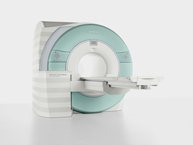
From Siemens Medical Systems;
Received FDA clearance in 2007.
The MAGNETOM Verio provides up to 102 integrated matrix coil elements and up to 32 independent radiofrequency channels that allow flexible coil combinations to make patient and coil repositioning virtually unnecessary. The Tim (total imaging matrix) technology also increases patient throughput due to a shorter scan time.
The open bore design offers great comfort for patients of all shapes and sizes.
Device Information and Specification
CLINICAL APPLICATION
Whole Body
CONFIGURATION
Ultra-short open bore
3 Tesla
Head, spine, torso/ body coil, neurovascular, cardiac, neck and multi-purpose flex coils. Peripheral vascular, breast, shoulder, knee, wrist, foot//ankle, TMJ optional.
CHANNELS (min. / max. configuration)
8, 18, 32
Chemical shift imaging, single voxel spectroscopy
IMAGING TECHNIQUES
iPAT, mSENSE and GRAPPA (image, k-space), noncontrast angiography, radial motion compensation, Dixon
MINIMUM TR
3-D GRE: 1.5 (256 matrix)
MINIMUM TE
3-D GRE: 0.63 (256 matrix)
FOV
0.5 - 50 cm
BORE DIAMETER
or W x H
At isocenter: L-R 70 cm, A-P 55 cm
TABLE CAPACITY
250 kg
MAGNET WEIGHT (gantry included)
8200 kg
DIMENSION H*W*D (gantry included)
173 x 230 x 222 cm
5-GAUSS FRINGE FIELD
2.6 m / 4.6 m
CRYOGEN USE
Zero boil off rate, refill approx. 10 years
COOLING SYSTEM
Water
up to 200 T/m/s
MAX. AMPLITUDE
45 mT/m
Passive, active; first order, second order standard
POWER REQUIREMENTS
380 / 400 / 420 / 440 / 460 / 480 V, 3-phase + ground; 110 kVA
spacer
MRI Resources 
Societies - Nerve Stimulator - Pacemaker - Knee MRI - MRI Technician and Technologist Jobs - Databases
 
MSK-Extremeâ„¢InfoSheet: - Devices -
Intro, 
Types of Magnets, 
Overview, 
etc.MRI Resource Directory:
 - Devices -
 
www.onicorp.com/ From ONI Medical Systems, Inc.;
MSK-Extremeâ„¢MRI system is a dedicated high field extremity imaging device, designed to provide orthopedic surgeons and other physicians with detailed diagnostic images of the foot, ankle, knee, hand, wrist and elbow, all with the clinical confidence and advantages derived from high field, whole body MRI units. The light weight (less than 650 kg) of the OrthOne System performs rapid patient studies, is easy to operate, has a patient friendly open environment and can be installed in a practice office or hospital, all at a cost similar to a low field extremity machine.
New features include a more powerful operating system that offers increased scan speed as well as a 160-mm knee coil with higher signal to noise ratio, and the option of a CD burner.
Device Information and Specification
CLINICAL APPLICATION
Dedicated extremity imaging
CONFIGURATION
16 cm knee, 18 cm lower extremity;; 12.3 cm upper extremity, additional high resolution v-SPEC Coils: 80 mm, 100 mm, or 145 mm.
SYNCHRONIZATION
No
PULSE SEQUENCES
SE, FSE, GE2D, GE3D, Inversion recovery (IR), Driven Equilibrium, Fat Saturation (FS), STIR, MT, PD, Flow Compensation (FC), RF spoiling, MTE, No Phase Wrap (NPW)
IMAGING MODES
Scout, single, multislice, volume
TR
10-10,000ms; 1ms steps
TE
5-150ms; 1 ms steps
SINGLE/MULTI SLICE
2D less than 200 msec/image
4cm-16cm
2D: 2mm-10mm/.1mm incr.
Up to 1,000x1,000
MEASURING MATRIX
X/Y: 64-512; 2 pixel steps
PIXEL INTENSITY
4,096 grey lvls; 256 lvls in 3D
28cm ID x 50cm L
MAGNET WEIGHT
635 kg
H*W*D
146 x 69 x 84 cm
POWER REQUIREMENTS
115VAC, 1phase, 20A; 208VAC, 3 phase, 30A
COOLING SYSTEM TYPE
LHe with 2 stage cold head
Negligible
STRENGTH
15 mT/m
5-GAUSS FRINGE FIELD
1.25m radial x 1.8m axial
Passive
spacer
 
Further Reading:
  Basics:
MSK Extreme Brochure(.pdf)
   by www.nova-logic.ch    
MSK Extreme Specifications(.pdf)
   by www.nova-logic.ch    
MRI Resources 
Spectroscopy - Quality Advice - Process Analysis - MRI Training Courses - Devices - Mobile MRI
 
Motion ArtifactInfoSheet: - Artifacts - 
Case Studies, 
Reduction Index, 
etc.MRI Resource Directory:
 - Artifacts -
 
Quick Overview
Please note that there are different common names for this artifact.
Artifact Information
NAME
Motion, phase encoded motion, instability, smearing
DESCRIPTION
Blurring and ghosting
REASON
Movement of the imaged object
HELP
Compensation techniques, more averages, anti spasmodic
Patient motion is the largest physiological effect that causes artifacts, often resulting from involuntary movements (e.g. respiration, cardiac motion and blood flow, eye movements and swallowing) and minor subject movements.
Movement of the object being imaged during the sequence results in inconsistencies in phase and amplitude, which lead to blurring and ghosting. The nature of the artifact depends on the timing of the motion with respect to the acquisition. Causes of motion artifacts can also be mechanical vibrations, cryogen boiling, large iron objects moving in the fringe field (e.g. an elevator), loose connections anywhere, pulse timing variations, as well as sample motion. These artifacts appear in the phase encoding direction, independent of the direction of the motion.
mri safety guidance
Image Guidance
Motion artifacts can be flipped 90° by swapping the phase//frequency encoding directions.
The artifacts can be reduced by using breath holding, cardiac synchronization or respiratory compensation techniques: triggering, gating, retrospective triggering or phase encoding artifact reduction. Flow effects can be reduced by using gradient moment nulling of the first order of flow, gradient moment rephasing or flow compensation, depending of the MRI system.
Peristaltic motion can be reduced with the intravenous injection of an anti-spasmodic (e.g. Buscopan).
By using multiple averages, respiratory motion can be reduced in the same way that multiple averages increase the signal to noise ratio. Noticeable motion averaging is seen when four averages are obtained, six averages are often as good as respiratory compensation techniques and higher averages will continue to improve image quality.
In some cases will help a presaturation of the anatomy that was generating the motion.

See also Phase Encoded Motion Artifact.
spacer

• View the DATABASE results for 'Motion Artifact' (24).Open this link in a new window

 
Further Reading:
  Basics:
The Effects of Breathing Motion on DCE-MRI Images: Phantom Studies Simulating Respiratory Motion to Compare CAIPIRINHA-VIBE, Radial-VIBE, and Conventional VIBE
Tuesday, 7 February 2017   by www.kjronline.org    
  News & More:
Patient movement during MRI: Additional points to ponder
Tuesday, 5 January 2016   by www.healthimaging.com    
Motion-compensation of Cardiac Perfusion MRI using a Statistical Texture Ensemble(.pdf)
June 2003   by www.imm.dtu.dk    
Searchterm 'Cryogen' was also found in the following services: 
spacer
News  (7)  Resources  (3)  Forum  (3)  
 
Panorama 0.23Tâ„¢InfoSheet: - Devices -
Intro, 
Types of Magnets, 
Overview, 
etc.MRI Resource Directory:
 - Devices -
 
www.medical.philips.com/main/products/mri/products/panoramafamily/panorama0.23t_rt/features/ From Philips Medical Systems;
the Panorama 0.23 T, providing a new design optimized for patient comfort, faster reconstruction time than before (300 images/second) and new gradient specifications. Philips' Panorama 0.23 T I/T supports MR-guided interventions, resulting in minimally invasive procedures, more targeted surgery, reduced recovery time and shorter hospital stays. Optional OptoGuide functionality enables real-time needle tracking. Philips' Panorama 0.23 TPanorama 0.2 R/T is the first and only open MRI system to enable radiation therapy planning using MR data sets. The Panorama also features the new and consistent Philips User Interface, an essential element of the Vequion clinical IT family of products and services.
Device Information and Specification
CLINICAL APPLICATION
Whole body
CONFIGURATION
Open MRI/C-arm
Head, head-neck, extremity M-L, neck, body/spine S-XL, shoulder, bilateral breast, wrist, TMJ, flex XS-S-M-L-XL-XXL
SYNCHRONIZATION
ECG/peripheral: Optional/optional, respiratory gating
PULSE SEQUENCES
SE, FE, IR, FFE, DEFFE, DESE, TSE, DETSE, Single shot SE, DRIVE, Balanced FFE, MRCP, Fluid Attenuated Inversion Recovery, Turbo FLAIR, IR-TSE, T1-STIR TSE, T2-STIR TSE, Diffusion Imaging, 3D SE, 3D FFE, MTC;; Angiography: CE-ANGIO, MRA 2D, 3D TOF
IMAGING MODES
Single, multislice, volume study, dynamic, SIMEX, multi chunk 3D, multiple stacks
TR
Min. 6.2 msec
TE
Min. 2.8 msec
SINGLE/MULTI SLICE
50 slices/sec
0.4 cm - 40 cm
1280 X 1024
MEASURING MATRIX
Up to 512 x 512
PIXEL INTENSITY
256 gray scale
MAGNET TYPE
Resistive/iron core
Open x 46 cm x infinite (side-first patient entry)
MAGNET WEIGHT
13110 kg
H*W*D
196 x 121 x 176 cm
POWER REQUIREMENTS
400/480 V
COOLING SYSTEM TYPE
Closed loop chilled water (chiller included)
N/A
STRENGTH
19 mT/m
5-GAUSS FRINGE FIELD
2.4 m / 3.7 m
Passive/active
spacer

• View the DATABASE results for 'Panorama 0.23T™' (2).Open this link in a new window

 
Further Reading:
  News & More:
Magnetic resonance imaging guided musculoskeletal interventions at 0.23T: Chapter 4. Materials and methods
2002
MRI Resources 
Spine MRI - Education - Movies - MRI Technician and Technologist Career - Quality Advice - Used and Refurbished MRI Equipment
 
Panorama 0.6TPanorama 0.2InfoSheet: - Devices -
Intro, 
Types of Magnets, 
Overview, 
etc.MRI Resource Directory:
 - Devices -
 
www.medical.philips.com/main/products/mri/products/panoramafamily/panorama0.23t_rt/features/ From Philips Medical Systems;
Panorama 0.6 T is the Philips Mid-Field Open MRI system. It is the most open MR scanner in the market, optimized for patient comfort and faster reconstruction time.
Device Information and Specification
CLINICAL APPLICATION
Whole body
CONFIGURATION
Open MRI/C-arm
Head, head-neck, extremity, neck, body/spine M-XL, shoulder, bilateral breast, wrist, TMJ, flex XS-S-M-L-XL-XXL
SYNCHRONIZATION
ECG/peripheral: Optional/yes, respiratory gating
PULSE SEQUENCES
SE, FE, IR, STIR, FFE, DEFFE, DESE, TSE, DETSE, Single shot SE, DRIVE, Balanced FFE, MRCP, Fluid Attenuated Inversion Recovery, Turbo FLAIR, IR-TSE, T1-STIR TSE, T2-STIR TSE, Diffusion Imaging, 3D SE, 3D FFE, Contrast Perfusion Analysis, MTC;; Angiography: CE-ANGIO, MRA 2D, 3D TOF
IMAGING MODES
Single, multislice, volume study, dynamic, SIMEX, multi chunk 3D, multiple stacks
TR
Min. 4.6 msec
TE
Min. 2.3 msec
SINGLE/MULTI SLICE
50 slices/sec
0.4 cm - 42 cm
1280 X 1024
MEASURING MATRIX
Up to 512 x 512
PIXEL INTENSITY
256 gray scale
MAGNET TYPE
Superconducting / iron core
Open x 47 cm x infinite (side-first patient entry)
MAGNET WEIGHT
38000 kg
H*W*D
254 x 244 x 325 cm
POWER REQUIREMENTS
400/480 V
COOLING SYSTEM TYPE
Liquid helium//air cool
0.00 L/hr helium
STRENGTH
20 mT/m
5-GAUSS FRINGE FIELD
2.4 m / 2.5 m
Passive/active
spacer

• View the DATABASE results for 'Panorama 0.6T™' (2).Open this link in a new window

MRI Resources 
IR - MRI Technician and Technologist Career - MRI Reimbursement - Services and Supplies - Process Analysis - MRI Accidents
 
previous      41 - 45 (of 61)     next
Result Pages : [1]  [2 3 4 5 6 7 8 9 10 11 12 13]
 Random Page
 
Share This Page
FacebookTwitterLinkedIn

MR-TIP    
Community   
User
Pass
Forgot your UserID/Password ?    


Next big thing in MRI will be :
AI 
remote operator 
personalized protocols 
helium-free 
molecular MRI 
portable MRI 

Look
      Ups


MR-TIP.com uses cookies! By browsing MR-TIP.com, you agree to our use of cookies.

Magnetic Resonance - Technology Information Portal
Member of SoftWays' Medical Imaging Group - MR-TIP • Radiology-TIP • Medical-Ultrasound-Imaging • 
Copyright © 2003 - 2024 SoftWays. All rights reserved. [ 19 April 2024]
Terms of Use | Privacy Policy | Advertising
 [last update: 2024-02-26 03:41:00]