| Info
Sheets |
| | | | | | | | | | | | | | | | | | | | | | | | |
 | Out-
side |
| | | | |
|
| | | | |
Result : Searchterm 'Cryogen' found in 2 terms [ ] and 59 definitions [ ] and 59 definitions [ ] ]
| | previous 6 - 10 (of 61) nextResult Pages : [1] [2 3 4 5 6 7 8 9 10 11 12 13] |  | |  | Searchterm 'Cryogen' was also found in the following services: | | | | |
| |  |
| |
|
Device Information and Specification
CLINICAL APPLICATION
Whole body
GRE, IR, FIR, STIR, TrueIR/FISP, FSE, FLAIR, MT, SS-FSE, MT-SE, MTC, MSE, EPI, GMR, fat/water sat./exc.
IMAGING MODES
Single, multislice, volume study, multi angle, multi oblique
TR
2.4 msec std.; 2.0 opt.; 1.8 w/ 30mT/m at 256matrix
TE
1.1 msec std.; 0.9 opt.; 0.78 w/30 mT/m at 256matrix
178 images/sec at 256 x 256 at 100% FOV
1024 x 1024 full screen display
21 micrometer in plane, 11 micrometer optional
MAGNET WEIGHT (gantry included)
3500kg, 5000kg in operation
DIMENSION H*W*D (gantry included)
POWER REQUIREMENTS
380/400/420/440/480 V
STRENGTH
20/35 mT/m standard, 30/52 opt.
Passive, act.; 1st order std./2nd opt.
| | | | | | | | | | |  Further Reading: Further Reading: | Basics:
|
|
| |
| | | | | |
| |
|
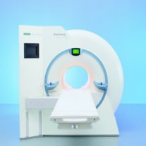
From Siemens Medical Systems;
while older navigator techniques take up to 40 minutes to create, the high performance of the MAGNETOM Sonata system enables 'complete examinations in less than 15 minutes'. It creates a new standard of diagnostic confidence and moves Cardiac MR from the research setting into routine clinical practice.
Device Information and Specification CLINICAL APPLICATION Whole body Body, head, spine, knee, neck, TMJ, extremity, head, breast, shoulder, others GRE, IR, FIR, STIR, TrueIR/FISP, FSE, FLAIR, MT, SS-FSE, MT-SE, MTC, MSE, EPI, 3D DESS//CISS/PSIF, GMR IMAGING MODES Single, multislice, volume study, multi angle, multi oblique178 images/sec at 256 x 256 at 100% FOV1024 x 1024 full screen display 4050kg, 5500kg in operation POWER REQUIREMENTS 380/400/420/440/480 V Passive, act.; 1st order std./2nd opt. | | | |
• View the DATABASE results for 'MAGNETOM Sonata™' (2).
| | | | |
| | | | | |
| |
|
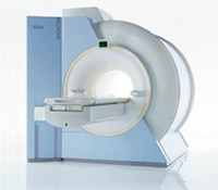
Device Information and Specification CLINICAL APPLICATION Whole body GRE, IR, FIR, STIR, TrueIR/FISP, FSE, FLAIR, MT, SS-FSE, MT-SE, MTC, MSE, EPI, GMR, fat/water sat./exc. IMAGING MODES Single, multislice, volume study, multi angle, multi obliqueTR 2.4 msec std.; 2.0 opt.; 1.8 w/30 mT/m at 256matrix TE 1.1 msec std.; 0.9 opt.; 0.78 w/30 mT/m at 256matrix 178 images/sec at 256 x 256 at 100% FOV1024 x 1024 full screen display 21 micrometer in plane, 11 micrometer optional 4050kg, 5500kg in operation H*W*D 236 x 215 x 160 cm w/covers POWER REQUIREMENTS 380/400/420/440/480 V STRENGTH 20/35 mT/m standard, 30/52 opt. Passive, act.; 1st order std./2nd opt. | | | |
• View the DATABASE results for 'MAGNETOM Symphony™' (2).
| | | | | | Further Reading: | Basics:
|
|
| |
| | | Searchterm 'Cryogen' was also found in the following services: | | | | |
| | |
| |
|
Magnetic resonance imaging ( MRI) is based on the magnetic resonance phenomenon, and is used for medical diagnostic imaging since ca. 1977 (see also MRI History).
The first developed MRI devices were constructed as long narrow tunnels. In the meantime the magnets became shorter and wider. In addition to this short bore magnet design, open MRI machines were created. MRI machines with open design have commonly either horizontal or vertical opposite installed magnets and obtain more space and air around the patient during the MRI test.
The basic hardware components of all MRI systems are the magnet, producing a stable and very intense magnetic field, the gradient coils, creating a variable field and radio frequency (RF) coils which are used to transmit energy and to encode spatial positioning. A computer controls the MRI scanning operation and processes the information.
The range of used field strengths for medical imaging is from 0.15 to 3 T. The open MRI magnets have usually field strength in the range 0.2 Tesla to 0.35 Tesla. The higher field MRI devices are commonly solenoid with short bore superconducting magnets, which provide homogeneous fields of high stability.
There are this different types of magnets:
The majority of superconductive magnets are based on niobium-titanium (NbTi) alloys, which are very reliable and require extremely uniform fields and extreme stability over time, but require a liquid helium cryogenic system to keep the conductors at approximately 4.2 Kelvin (-268.8° Celsius). To maintain this temperature the magnet is enclosed and cooled by a cryogen containing liquid helium (sometimes also nitrogen).
The gradient coils are required to produce a linear variation in field along one direction, and to have high efficiency, low inductance and low resistance, in order to minimize the current requirements and heat deposition. A Maxwell coil usually produces linear variation in field along the z-axis; in the other two axes it is best done using a saddle coil, such as the Golay coil.
The radio frequency coils used to excite the nuclei fall into two main categories; surface coils and volume coils.
The essential element for spatial encoding, the gradient coil sub-system of the MRI scanner is responsible for the encoding of specialized contrast such as flow information, diffusion information, and modulation of magnetization for spatial tagging.
An analog to digital converter turns the nuclear magnetic resonance signal to a digital signal. The digital signal is then sent to an image processor for Fourier transformation and the image of the MRI scan is displayed on a monitor.
For Ultrasound Imaging (USI) see Ultrasound Machine at Medical-Ultrasound-Imaging.com.
See also the related poll results: ' In 2010 your scanner will probably work with a field strength of' and ' Most outages of your scanning system are caused by failure of' | | | | | | | | |
• View the DATABASE results for 'Device' (141).
| | |
• View the NEWS results for 'Device' (29).
| | | | | | Further Reading: | News & More:
|
 |
small-steps-can-yield-big-energy-savings-and-cut-emissions-mris
Thursday, 27 April 2023 by www.itnonline.com | | |
Portable MRI can detect brain abnormalities at bedside
Tuesday, 8 September 2020 by news.yale.edu | | |
Point-of-Care MRI Secures FDA 510(k) Clearance
Thursday, 30 April 2020 by www.diagnosticimaging.com | | |
World's First Portable MRI Cleared by FDA
Monday, 17 February 2020 by www.medgadget.com | | |
Low Power MRI Helps Image Lungs, Brings Costs Down
Thursday, 10 October 2019 by www.medgadget.com | | |
Cheap, portable scanners could transform brain imaging. But how will scientists deliver the data?
Tuesday, 16 April 2019 by www.sciencemag.org | | |
The world's strongest MRI machines are pushing human imaging to new limits
Wednesday, 31 October 2018 by www.nature.com | | |
Kyoto University and Canon reduce cost of MRI scanner to one tenth
Monday, 11 January 2016 by www.electronicsweekly.com | | |
A transportable MRI machine to speed up the diagnosis and treatment of stroke patients
Wednesday, 22 April 2015 by medicalxpress.com | | |
Portable 'battlefield MRI' comes out of the lab
Thursday, 30 April 2015 by physicsworld.com | | |
Chemists develop MRI technique for peeking inside battery-like devices
Friday, 1 August 2014 by www.eurekalert.org | | |
New devices doubles down to detect and map brain signals
Monday, 23 July 2012 by scienceblog.com |
|
| |
| | | | | |
| |
|
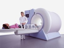
Device Information and Specification
CLINICAL APPLICATION
Whole body
SE, IR, FSE, FIR, GE, SG, BASG, PBSG, PCIR, DWI, Radial, Angiography: TOF, FLUTE (Fluoro-triggered bolus MRA), Time-resolved MRA
IMAGING MODES
Single, multislice, volume study
Level Range: -2,000 to +4,000
POWER REQUIREMENTS
208/220/240 V, single phase
| | | |
• View the DATABASE results for 'Echelon™ 1.5T' (2).
| | |
• View the NEWS results for 'Echelon™ 1.5T' (3).
| | | | | | Further Reading: | Basics:
|
|
| |
| | | | |
| | |
| | | |
|
| |
 | Look
Ups |
| |