 | Info
Sheets |
| | | | | | | | | | | | | | | | | | | | | | | | |
 | Out-
side |
| | | | |
|
| | | | | |  | Searchterm 'Coil' was also found in the following services: | | | | |
|  |  |
| |
|
(SENSE) A MRI technique for relevant scan time reduction. The spatial information related to the coils of a receiver array are utilized for reducing conventional Fourier encoding. In principle, SENSE can be applied to any imaging sequence and k-space trajectories. However, it is particularly feasible for Cartesian sampling schemes. In 2D Fourier imaging with common Cartesian sampling of k-space sensitivity encoding by means of a receiver array enables to reduce the number of Fourier encoding steps.
SENSE reconstruction without artifacts relies on accurate knowledge of the individual coil sensitivities. For sensitivity assessment, low-resolution, fully Fourier-encoded reference images are required, obtained with each array element and with a body coil.
The major negative point of parallel imaging techniques is that they diminish SNR in proportion to the numbers of reduction factors.
R is the factor by which the number of k-space samples is reduced. In standard Fourier imaging reducing the sampling density results in the reduction of the FOV, causing aliasing. In fact, SENSE reconstruction in the Cartesian case is efficiently performed by first creating one such aliased image for each array element using discrete Fourier transformation (DFT).
The next step then is to create a full-FOV image from the set of intermediate images. To achieve this one must undo the signal superposition underlying the fold-over effect. That is, for each pixel in the reduced FOV the signal contributions from a number of positions in the full FOV need to be separated. These positions form a Cartesian grid corresponding to the size of the reduced FOV.
The advantages are especially true for contrast-enhanced MR imaging such as
dynamic liver MRI (liver imaging) ,
3 dimensional magnetic resonance angiography (3D MRA), and magnetic resonance cholangiopancreaticography ( MRCP).
The excellent scan speed of SENSE allows for acquisition of two separate sets of hepatic MR images within the time regarded as the hepatic arterial-phase (double arterial-phase technique) as well as that of multidetector CT.
SENSE can also increase the time efficiency of spatial signal encoding in 3D MRA. With SENSE, even ultrafast (sub second) 4D MRA can be realized.
For MRCP acquisition, high-resolution 3D MRCP images can be constantly provided by SENSE. This is because SENSE resolves the presence of the severe motion artifacts due to longer acquisition time. Longer acquisition time, which results in diminishing image quality, is the greatest problem for 3D MRCP imaging.
In addition, SENSE reduces the train of gradient echoes in combination with a faster k-space traversal per unit time, thereby dramatically improving the image quality of single shot echo planar imaging (i.e. T2 weighted, diffusion weighted imaging). | | | | | | | | | | |  Further Reading: Further Reading: | News & More:
|
|
| |
| | | Searchterm 'Coil' was also found in the following services: | | | | |
| | |
| |
|
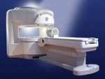
From GE Healthcare;
the Signa Ovation™ is a patient-friendly open MRI scanner designed not only to handle a typical patient mix, but to accommodate larger patients, patients who are claustrophobic, and others who have difficulty tolerating the close quarters of conventional MR machines.
Device Information and Specification CLINICAL APPLICATION Whole body Standard: SE, IR, 2D/3D GRE and SPGR, 2D/3D TOF, 2D/3D FSE, 2D/3D FGRE and FSPGR, SSFP, FLAIR, EPI, optional: 2D/3D Fiesta, true chem sat, fat/water separation, single shot diffusion EPI, line scan diffusionIMAGING MODES Localizer, single slice, multislice, volume, fast, POMP, multi slab, cine, slice and frequency zip, extended dynamic range, tailored RF TR 1.3 to 12000 msec in increments of 1 msec TE 0.4 to 2000 msec in increments of 1 msec 2D: 1.4mm - 20mm 3D: 0.2mm - 20mm 0.08 mm; 0.02 mm optional POWER REQUIREMENTS 200 - 480, 3-phase MAX. GRADIENT AMPLITUDE 19 mT/m | | | |
• View the DATABASE results for 'Signa Ovation™' (2).
| | | | |
| | | | | |
| |
|
Time-dependent electromagnetic fields are significantly attenuated by conducting media (including the human body); the skin depth gives a measure of the average depth of penetration of the RF field.
A high power frequency tunable RF source can be rapidly switched on and off. This produces a large RF field perpendicular to the magnetic field. This RF field is focused by the body coil. The RF source and coils must be tunable in both frequency and impedance to
'match the impedance' of the patient's body.
The skin depth may be a limiting factor in MR imaging at very high frequencies (high magnetic fields). The skin depth also affects the Q of the coils. | | | | | | | Further Reading: | News & More:
|
|
| |
| | | Searchterm 'Coil' was also found in the following services: | | | | |
| | |
| |
|

From Hitachi Medical Systems America Inc.;
the AIRIS II, an entry in the diagnostic category of open MR systems, was designed by Hitachi
Medical Systems America Inc. (Twinsburg, OH, USA) and Hitachi Medical Corp. (Tokyo) and is manufactured by the Tokyo branch. A 0.3 T field-strength magnet and phased array coils deliver high image quality without the need for a tunnel-type high-field system, thereby significantly improving patient comfort not only for claustrophobic patients.
Device Information and Specification
CLINICAL APPLICATION
Whole body
QD Head, MA Head and Neck, QD C-Spine, MA or QD Shoulder, MA CTL Spine, QD Knee, Neck, QD TMJ, QD Breast, QD Flex Body (4 sizes), Small and Large Extrem., QD Wrist, MA Foot and Ankle (WIP), PVA (WIP)
SE, GE, GR, IR, FIR, STIR, FSE, ss-FSE, FLAIR, EPI -DWI, SE-EPI, ms - EPI, SSP, MTC, SARGE, RSSG, TRSG, MRCP, Angiography: CE, 2D/3D TOF
IMAGING MODES
Single, multislice, volume study
TR
SE: 30 - 10,000msec GE: 20 - 10,000msec IR: 50 - 16,700msec FSE: 200 - 16,7000msec
TE
SE : 10 - 250msec IR: 10 -250msec GE: 5 - 50 msec FSE: 15 - 2,000
0.05 sec/image (256 x 256)
2D: 2 - 100 mm; 3D: 0.5 - 5 mm
Level Range: -2,000 to +4,000
POWER REQUIREMENTS
208/220/240 V, single phase
COOLING SYSTEM TYPE
Air-cooled
2.0 m lateral, 2.5 m vert./long
| | | |
• View the DATABASE results for 'AIRIS II™' (2).
| | | | |
| | | Searchterm 'Coil' was also found in the following services: | | | | |
| | |
| |
|
Quick Overview
Please note that there are different common names for this MRI artifact.
DESCRIPTION
Image wrap around
Aliasing is an artifact that occurs in MR images when the scanned body part is larger than field of view ( FOV). As a consequence of the acquired k-space frequencies not being sampled densely enough, whereby portions of the object outside of the desired FOV get mapped to an incorrect location inside the FOV. The cyclical property of the Fourier transform fills the missing data of the right side with data from behind the FOV of the left side and vice versa. This is caused by a too small number of samples acquired in, e.g. the frequency encoding direction, therefore the spectrums will overlap, resulting in a replication of the object in the x direction.
Aliasing in the frequency direction can be eliminated by twice as fast sampling of the signal or by applying frequency specific filters to the received signal.
A similar problem occurs in the phase encoding direction, where the phases of signal-bearing tissues outside of the FOV in the y-direction are a replication of the phases that are encoded within the FOV. Phase encoding gradients are scaled for the field of view only, therefore tissues outside the FOV do not get properly phase encoded relative to their actual position and 'wraps' into the opposite side of the image.

Image Guidance
| | | |
• View the DATABASE results for 'Aliasing Artifact' (11).
| | | | |
| | | | |
| | |
| | | |
|
| |
 | Look
Ups |
| |


