 | Info
Sheets |
| | | | | | | | | | | | | | | | | | | | | | | | |
 | Out-
side |
| | | | |
|
| | | | |
Result : Searchterm 'Cine' found in 5 terms [ ] and 52 definitions [ ] and 52 definitions [ ] ]
| previous 21 - 25 (of 57) nextResult Pages :  [1] [1]  [2 3 4 5 6 7 8 9 10 11 12] [2 3 4 5 6 7 8 9 10 11 12] |  | |  | Searchterm 'Cine' was also found in the following services: | | | | |
|  |  |
| |
|
Ferric ammonium citrate is a complex salt of indefinite composition that contains varying amounts of iron, that is obtained as red crystals or a brownish yellow powder or as green crystals or powder, and that was used formerly in medi cine for treating iron-deficiency anaemia.
Solutions (e.g. corn oil emulsion) of ferric ammonium citrate (e.g., FerriSeltz, Geritol) can be used as positive oral contrast agents in MRI. Ferric ammonium citrate is safe and effective in humans, but has minor side effects.
See also Classifications, Characteristics, etc. | |  | | | |
|  |  | Searchterm 'Cine' was also found in the following services: | | | | |
|  |  |
| |
|
Short name: Ami 227, generic name: Ferumoxtran, (USPIO)
Ferumoxtran is a substance of the class of ultrasmall superparamagnetic iron oxide used as a lymph node specific contrast agent for MRI.
See also Combidex®, Sinerem® and Ultrasmall Superparamagnetic Iron Oxide.
Partner(s): Cytogen Corporation, National Cancer Institute.
An approvable letter was received from the U.S. Food and Drug Administration for Combidex in June 2000. Advanced Magnetics, Inc. has submitted a complete response to the approvable letter received from the U.S. Food and Drug Administration, which was accepted by the FDA and assigned a user fee goal date of March 30, 2005.
In Europe, a Dossier (the European equivalent of a NDA) was submitted by Advanced Magnetics' European partner, Guerbet SA, to the European Medi cines Evaluations Agency in December 1999.
( Sinerem® is the brand name for this USPIO in Europe manufactured by Guerbet, Combidex® by Advanced Magnetics for the U.S. market)
Advanced Magnetics, Inc. changed its name in July 2007 to AMAG Pharmaceuticals Inc. | |  | |
• View the DATABASE results for 'Ferumoxtran' (3).
| | | | |  Further Reading: Further Reading: | | Basics:
|
|
News & More:
| |
| |
|  | |  |  |  |
| |
|
Flow phenomena are intrinsic processes in the human body. Organs like the heart, the brain or the kidneys need large amounts of blood and the blood flow varies depending on their degree of activity. Magnetic resonance imaging has a high sensitivity to flow and offers accurate, reproducible, and noninvasive methods for the quantification of flow. MRI flow measurements yield information of blood supply of of various vessels and tissues as well as cerebro spinal fluid movement.
Flow can be measured and visualized with different pulse sequences (e.g. phase contrast sequence, cine sequence, time of flight angiography) or contrast enhanced MRI methods (e.g. perfusion imaging, arterial spin labeling).
The blood volume per time (flow) is measured in: cm3/s or ml/min. The blood flow-velocity decreases gradually dependent on the vessel diameter, from approximately 50 cm per second in arteries with a diameter of around 6 mm like the carotids, to 0.3 cm per second in the small arterioles.
Different flow types in human body:
•
Behaves like stationary tissue, the signal intensity depends on T1, T2 and PD = Stagnant flow
•
Flow with consistent velocities across a vessel = Laminar flow
•
Laminar flow passes through a stricture or stenosis (in the center fast flow, near the walls the flow spirals) = Vortex flow
•
Flow at different velocities that fluctuates = Turbulent flow
See also Flow Effects, Flow Artifact, Flow Quantification, Flow Related Enhancement, Flow Encoding, Flow Void, Cerebro Spinal Fluid Pulsation Artifact, Cardiovascular Imaging and Cardiac MRI. | | | |  | |
• View the DATABASE results for 'Flow' (113).
| | |
• View the NEWS results for 'Flow' (7).
| | | | |  Further Reading: Further Reading: | News & More:
|
|
| |
|  |  | Searchterm 'Cine' was also found in the following services: | | | | |
|  |  |
| |
|

From Esaote S.p.A.;
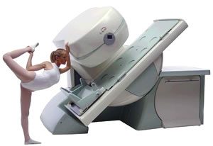
Esaote introduced the new G-SCAN at the RSNA in Dec. 2004. The G-SCAN covers almost all musculoskeletal applications including the spine. The tilting gantry is designed for scanning in weight-bearing positions. This unique MRI scanner is developed in line with the Esaote philosophy of creating high quality MRI systems that are easy to install and that have a low breakeven point.
Device Information and Specification
SE, GE, IR, STIR, TSE, 3D CE, GE-STIR, 3D GE, ME, TME, HSE
100 up to 350 mm, 25 mm displayed
POWER REQUIREMENTS
100/110/200/220/230/240 V
| |  | |
• View the DATABASE results for 'G-SCAN' (3).
| | | | |
|  |  | Searchterm 'Cine' was also found in the following services: | | | | |
|  | |  | |  |  |
|  | | |
|
| |
 | Look
Ups |
| |