 | Info
Sheets |
| | | | | | | | | | | | | | | | | | | | | | | | |
 | Out-
side |
| | | | |
|
| | | | |
Result : Searchterm 'Cine' found in 5 terms [ ] and 52 definitions [ ] and 52 definitions [ ] ]
| | previous 51 - 55 (of 57) nextResult Pages : [1] [2 3 4 5 6 7 8 9 10 11 12] |  | |  | Searchterm 'Cine' was also found in the following services: | | | | |
| |  |
| |
|
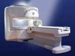
From GE Healthcare;
the Signa Ovation™ is a patient-friendly open MRI scanner designed not only to handle a typical patient mix, but to accommodate larger patients, patients who are claustrophobic, and others who have difficulty tolerating the close quarters of conventional MR machines.
Device Information and Specification CLINICAL APPLICATION Whole body Standard: SE, IR, 2D/3D GRE and SPGR, 2D/3D TOF, 2D/3D FSE, 2D/3D FGRE and FSPGR, SSFP, FLAIR, EPI, optional: 2D/3D Fiesta, true chem sat, fat/water separation, single shot diffusion EPI, line scan diffusionIMAGING MODES Localizer, single slice, multislice, volume, fast, POMP, multi slab, cine, slice and frequency zip, extended dynamic range, tailored RF TR 1.3 to 12000 msec in increments of 1 msec TE 0.4 to 2000 msec in increments of 1 msec 2D: 1.4mm - 20mm 3D: 0.2mm - 20mm 0.08 mm; 0.02 mm optional POWER REQUIREMENTS 200 - 480, 3-phase MAX. GRADIENT AMPLITUDE 19 mT/m | | | | | |
| | | Searchterm 'Cine' was also found in the following services: | | | | |
| | |
| |
|
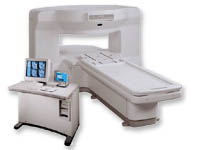
From GE Healthcare;
the New Signa Profile/i is a patient friendly open MRI system that virtually eliminates patient anxiety and claustrophobia, without compromising diagnostic utility.
Device Information and Specification CLINICAL APPLICATION Whole body Integrated transmit body coil, body flex sizes: M, L, XL, quadrature, head coil quadrature, 4 channel neurovascular array, 8 channel CTL array, quad. c- spine, 2 channel shoulder array, extremity coil, 3 channel wrist array, 4 channel breast array, 6, 9, 11 inch general purpose loop coils Standard: SE, IR, 2D/3D GRE and SPGR, Angiography: 2D/3D TOF, 2D/3D phase contrast; 2D/3D FSE, 2D/3D FRFSE, FGRE and FSPGR, SSFP, FLAIR, EPI, optional: 2D/3D Fiesta, fat/water separation, T1 FLAIRIMAGING MODES Localizer, single slice, multislice, volume, fast, POMP, multi slab, cine, slice and frequency zip, extended dynamic range, tailored RF TR 6 to 12000 msec in increments of 1 msec TE 1.3 to 2000 msec in increments of 1 msec 2D: 2.7mm - 20mm 3D: 0.2mm - 5mm 0.08 mm; 0.02 mm optional 10,000 kg w/gradient enclosure POWER REQUIREMENTS 200 - 480, 3-phase COOLING SYSTEM TYPE None required | | | |
• View the DATABASE results for 'Signa Profile™' (2).
| | | | |
| | | | | |
| |
|

From GE Healthcare;
The Signa SP 0.5T™ is an open MRI magnet that is designed for use in interventional radiology and intra-operative imaging. The vertical gap configuration increases patient positioning options, improves patient observation, and allows continuous access to the patient during imaging.
The magnet enclosure also incorporates an intercom, patient observation video camera, laser patient alignment lights, and task lighting in the imaging volume.
Device Information and Specification CLINICAL APPLICATION Whole body Integrated transmit and receive body coil; optional rotational body coil, head; other coils optional; open architecture makes system compatible with a wide selection of coilsarray Standard: SE, IR, 2D/3D GRE and SPGR, 2D/3D TOF, 2D/3D FSE, 2D/3D FGRE and FSPGR, SSFP, FLAIR, EPI, optional: 2D/3D Fiesta, true chem sat, fat/water separation, single shot diffusion EPI IMAGING MODES Localizer, single slice, multislice, volume, fast, POMP, multi slab, cine, slice and frequency zip, extended dynamic range, tailored RF TR 1.3 to 12000 msec in increments of 1 msec TE 0.4 to 2000 msec in increments of 1 msec 2D: 1.4mm - 20mm 3D: 0.2mm - 20mm POWER REQUIREMENTS 200 - 480, 3-phase | | | |
• View the DATABASE results for 'Signa SP 0.5T™ Open Configuration' (2).
| | | | |  Further Reading: Further Reading: | News & More:
|
|
| |
| | | Searchterm 'Cine' was also found in the following services: | | | | |
| | |
| |
|
For the wide uses of NMR spectroscopy (from mineralogy to medi cine) there is a variety of different spectroscopic imaging techniques available.
A short listing of the most frequent variations:
•
'Two-dimensional NMR Spectroscopy' (2D NMR) is based on pulse spectroscopy. This technique is mostly used for the study of chemical interactions accompanied by magnetization transfer. Examples for more diversified spectroscopy techniques are based on homonuclear (COSY, TOCSY, 2D-INADEQUATE, NOESY, ROESY) or heteronuclear correlation (HSQC, HMQC, HMBC).
•
'Solid State NMR Spectroscopy' analyzes samples with little or no molecular mobility. Dipolar coupling and chemical shift anisotropy are the dominating nuclear physical effects here. Used for example in pharmaceutical analysis.
•
'Solution State NMR Spectroscopy' is a technique to analyze the structure of samples with a high degree of molecular mobility as polymers, proteins, nucleic acids etc.
| | | |
• View the DATABASE results for 'Spectroscopic Imaging Techniques' (2).
| | | | | | Further Reading: | | Basics:
|
|
News & More:
| |
| |
| | | Searchterm 'Cine' was also found in the following services: | | | | |
| | |
| |
|
Edward Purcell and Felix Bloch discovered the basic of spectroscopy in 1946 (see MRI History). Nuclear magnetic resonance spectroscopy ( NMR Spectroscopy or MRS) is an analytical tool, based on nuclei that have a spin (nuclei with an odd number of neutrons and/or protons) like 1H, 13C, 17O, 19F, 31P etc.
Through nuclear magnetic principles as precession, chemical shift, spin spin coupling etc., the analysis of the content, purity, and molecular structure of a sample is possible. The spectrum produced by this process contains a number of peaks; the highs and the positions of these peaks allow the exact analysis. Unknown compounds can be matched against spectral libraries. Even very complex organic compounds as enzymes and proteins can be determined. For the wide uses of NMR spectroscopy (from mineralogy to medi cine) there is a variety of different techniques available.
See Spectroscopic Imaging Techniques. | | | |
• View the DATABASE results for 'Spectroscopy' (90).
| | |
• View the NEWS results for 'Spectroscopy' (3).
| | | | | | Further Reading: | | Basics:
|
|
News & More:
| |
| |
| | | | |
| | | |
|
| |
 | Look
Ups |
| |


