Magnetic Resonance - Technology Information Portal Welcome to MRI Technology
Info
  Sheets

Out-
      side


 'Cardiac Motion Artifact' 
SEARCH FOR    
 
  2 3 5 A B C D E F G H I J K L M N O P Q R S T U V W X Y Z
Result : Searchterm 'Cardiac Motion Artifact' found in 1 term [] and 1 definition [], (+ 10 Boolean[] results
1 - 5 (of 12)     next
Result Pages : [1]  [2 3]
MRI Resources 
MRI Technician and Technologist Schools - IR - Online Books - Resources - Abdominal Imaging - Collections
 
Cardiac Motion ArtifactInfoSheet: - Artifacts - 
Case Studies, 
Reduction Index, 
etc.MRI Resource Directory:
 - Artifacts -
 
Quick Overview
Artifact Information
NAME
Cardiac motion
DESCRIPTION
Blurring, ghosting
REASON
Cardiac motion
HELP
cardiac and respiratory synchronization
Movement of the heart causes blurring and ghosting in the images. The artifacts appear in the phase encoding direction, independent of the direction of the motion.
mri safety guidance
Image Guidance
These artifacts can be reduced by using cardiac synchronization: triggering, gating or retrospective triggering. Maximum reduction can be achieved by using triggering in combination with flow compensation, respiratory triggering or breath hold and regional saturation techniques.

See also Motion Artifact.
 
Images, Movies, Sliders:
 Cardiac Infarct Short Axis Cine bFFE 1  Open this link in a new window
    
 Normal Dual Inversion Fast Spin-echo  Open this link in a new window
 
spacer
 
• Share the entry 'Cardiac Motion Artifact':  Facebook  Twitter  LinkedIn  
 
Further Reading:
  Basics:
Motion-compensation of Cardiac Perfusion MRI using a Statistical Texture Ensemble(.pdf)
June 2003   by www.imm.dtu.dk    
  News & More:
Controlling patient's breathing makes cardiac MRI more accurate
Friday, 13 May 2016   by www.upi.com    
MRI Resources 
MRI Training Courses - MRI Accidents - Image Quality - Online Books - Education pool - Contrast Agents
 
Ghosting ArtifactInfoSheet: - Artifacts - 
Case Studies, 
Reduction Index, 
etc.MRI Resource Directory:
 - Artifacts -
 
Quick Overview
Artifact Information
NAME
Ghosting, ghost
DESCRIPTION
Displaced reduplications of image in phase encoding direction
REASON
Motion, heartbeat, respiration
HELP
Triggering, breath hold, pharmaceuticals to reduce bowel motion
Ghosting artifacts are in the most cases caused by movements (e.g., respiratory motion, bowel motion, arterial pulsations, swallowing, and heartbeat) and appear in the phase encoding direction.
mri safety guidance
Image Guidance
Ghosting artifacts can be reduced by respiratory and cardiac triggering, the use of breath holding pulse sequences, flow compensation or presaturation pulses, depending on their origin. To reduce bowel motion also pharmaceuticals, such as glucagon or scopolamine are useful. This will decrease artifacts from both peristalsis and breathing.

See also Motion Artifact, Phase Encoded Motion Artifact, Cardiac Motion Artifact, and Artifact Reduction - Motion.
spacer

• View the DATABASE results for 'Ghosting Artifact' (5).Open this link in a new window

 
Further Reading:
  Basics:
MRI Artifact Gallery
   by chickscope.beckman.uiuc.edu    
MRI Resources 
Safety pool - Portals - Spectroscopy - Intraoperative MRI - Used and Refurbished MRI Equipment - Databases
 
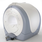
Signa HDx 3.0T™InfoSheet: - Devices -
Intro, 
Types of Magnets, 
Overview, 
etc.
 
gehealthcare.com/euen/mri/products/signa-hdx-3t/index.html From GE Healthcare;
The Signa HDx MRI system is GE's leading edge whole body magnetic resonance scanner designed to support high resolution, high signal to noise ratio, and short scan times.
Signa HDx 3.0T offers new technologies like ultra-fast image reconstruction through the new XVRE recon engine, advancements in parallel imaging algorithms and the broadest range of premium applications. The HD applications, PROPELLER (high-quality brain imaging extremely resistant to motion artifacts), TRICKS (contrast-enhanced angiographic vascular lower leg imaging), VIBRANT (for breast MRI), LAVA (high resolution liver imaging with shorter breath holds and better organ coverage) and MR Echo (high-definition cardiac images in real time) offer unique capabilities.
Device Information and Specification
CLINICAL APPLICATION
Whole body
CONFIGURATION
Compact short bore
Head and body coil, T/R quadrature head; optional coils e.g., T/R phased array extremity abdomen, spine, breast, knee, shoulder, cardiac imaging coils
SYNCHRONIZATION
ECG/peripheral, respiratory gating
PULSE SEQUENCES
SE, IR, 2D/3D GRE, RF-spoiled GRE, 2DFGRE, 2DFSPGR, 3DFGRE, 3DFSPGR, 3DTOFGRE, 3DFSPGR, 2DFSE, 2DFSE-XL, 2DFSE-IR, T1-FLAIR, SSFSE, EPI, DW-EPI, BRAVO, Angiography: 2D/3D TOF, 2D/3D phase contrast vascular
IMAGING MODES
Single, multislice, volume study, fast scan, multi slab, cine, localizer
1 cm to 40 cm continuous
2D 0.5 mm; 3D 0.1 mm
1024 x 1024
PIXEL INTENSITY
256 gray levels
60 cm
MAGNET WEIGHT
12000 kg
H*W*D
240 x 2216,6 x 201,6 cm
POWER REQUIREMENTS
480 or 380/415, 3 phase ||
COOLING SYSTEM TYPE
Closed-loop water-cooled grad.
0.03 L/hr helium
STRENGTH
23 - 50 mT/m
80 - 150 mT/m/ms
5-GAUSS FRINGE FIELD
2.8 m / 5.0 m
second and high order
spacer
MRI Resources 
Directories - Safety pool - Spectroscopy pool - Brain MRI - PACS - Calculation
 
ArtifactForum -
related threadsInfoSheet: - Artifacts - 
Case Studies, 
Reduction Index, 
etc.MRI Resource Directory:
 - Artifacts -
 
An image artifact is a structure not normally present but visible as a result of a limitation or malfunction in the hardware or software of the MRI device, or in other cases a consequence of environmental influences as heat or humidity or it can be caused by the human body (blood flow, implants etc.). The knowledge of MRI artifacts (brit. artefacts) and noise producing factors is important for continuing maintenance of high image quality. Artifacts may be very noticeable or just a few pixels out of balance but can give confusing artifactual appearances with pathology that may be misdiagnosed.
Changes in patient position, different pulse sequences, metallic artifacts, or other imaging variables can cause image distortions, which can be reduced by the operator; artifacts due to the MR system may require a service engineer.
Many types of artifacts may occur in magnetic resonance imaging. Artifacts in magnetic resonance imaging are typically classified as to their basic principles, e.g.:
Physiologic (motion, flow)
Hardware (electromagnetic spikes, ringing)
Inherent physics (chemical shift, susceptibility, metal)

Several techniques are developed to reduce these artifacts (e.g. respiratory compensation, cardiac gating, eddy current compensation) but sometimes these effects can also be exploited, e.g. for flow measurements.

See also the related poll result: 'Most outages of your scanning system are caused by failure of'
spacer

• View the DATABASE results for 'Artifact' (166).Open this link in a new window

 
Further Reading:
  Basics:
ARTEFACT VERSUS ARTIFACT
Saturday, 26 January 2002   by www.worldwidewords.org    
  News & More:
MRI results affected by movement? MIT researchers have an AI-powered solution
Friday, 25 August 2023   by healthimaging.com    
Magnetic eyelashes: A new source of MRI artifacts
Wednesday, 24 July 2019   by medicalxpress.com    
On the Horizon - Next Generation MRI
Wednesday, 23 October 2013   by thefutureofthings.com    
MRI Resources 
MRI Technician and Technologist Schools - Education pool - Intraoperative MRI - Implant and Prosthesis - Guidance - Pathology
 
Lung ImagingMRI Resource Directory:
 - Lung Imaging -
 
Lung imaging is furthermore a challenge in MRI because of the predominance of air within the lungs and associated susceptibility issues as well as low signal to noise of the inflated lung parenchyma. Cardiac and respiratory triggered or breath hold sequences allow diagnostic imaging, however a comparable image quality with computed tomography is still difficult to achieve.
Assumptions for lung MRI:
Low signal to noise ratio of the inherently low lung proton density.
Cardiac and respiratory motion artifacts.
Magnetic susceptibility effects of large magnetic field gradients.
Very short transverse relaxation times and significant diffusion yielding short T2 (30-70 msec), short T2* (1-3 msec), and additional long T1 relaxation times (1300-1500 msec).
The extreme short T2 values are responsible for a fast signal decay during a single shot readout, resulting in blurring.

The current trends in MRI are the use of new imaging technologies and increasingly powerful magnetic fields. Among these technologies are parallel imaging techniques as well as ventilation agents like hyperpolarized helium for the use as an inert inhalational contrast agent to study lung ventilation properties. With hyperpolarized gases clear images of the lungs can be obtained without using a large magnetic field (see also back projection imaging). Single shot sequences (e.g. TSE or Half Fourier Acquisition Single Shot Turbo Spin Echo HASTE) used in lung MR imaging benefits from parallel imaging techniques due to reduced relaxation time effects during the echo train and therefore reduced image blurring as well as reduced motion artifacts.
In the future, more effective contrast agents may provide an alternative solution to the need for high field MRI. Dynamic contrast enhanced MRI perfusion has demonstrated a potential in the diagnosis of pulmonary embolism or to characterize lung cancer and mediastinal tumors. 3D contrast enhanced magnetic resonance angiography of the thoracic vessel.

See also the related poll result: 'MRI will have replaced 50% of x-ray exams by'
 
Images, Movies, Sliders:
 Anatomic Imaging of the Lungs  Open this link in a new window
      

Courtesy of  Robert R. Edelman
 Normal Lung Gd Perfusion MRI  Open this link in a new window
      

Courtesy of  Robert R. Edelman

 MRI Thorax Basal Plane  Open this link in a new window
 
Radiology-tip.comradLung Scintigraphy
spacer

• View the DATABASE results for 'Lung Imaging' (7).Open this link in a new window


• View the NEWS results for 'Lung Imaging' (3).Open this link in a new window.
 
Further Reading:
  Basics:
A safer approach for diagnostic medical imaging
Monday, 29 September 2014   by www.eurekalert.org    
Parallel Lung Imaging(.pdf)
  News & More:
Chest MRI a viable alternative to chest CT in COVID-19 pneumonia follow-up
Monday, 21 September 2020   by www.healthimaging.com    
CT Imaging Features of 2019 Novel Corona virus (2019-nCoV)
Tuesday, 4 February 2020   by pubs.rsna.org    
Polarean Imaging Phase III Trial Results Point to Potential Improvements in Lung Imaging
Wednesday, 29 January 2020   by www.diagnosticimaging.com    
Low Power MRI Helps Image Lungs, Brings Costs Down
Thursday, 10 October 2019   by www.medgadget.com    
Chest MRI Using Multivane-XD, a Novel T2-Weighted Free Breathing MR Sequence
Thursday, 11 July 2019   by www.sciencedirect.co    
Researchers Review Importance of Non-Invasive Imaging in Diagnosis and Management of PAH
Wednesday, 11 March 2015   by lungdiseasenews.com    
New MRI Approach Reveals Bronchiectasis' Key Features Within the Lung
Thursday, 13 November 2014   by lungdiseasenews.com    
MRI techniques improve pulmonary embolism detection
Monday, 19 March 2012   by medicalxpress.com    
  News & More:
Partnership with VIDA to streamline adoption of advanced MRI of the lungs
Monday, 11 September 2023   by www.itnonline.com    
MRI Resources 
MR Myelography - Patient Information - Anatomy - Societies - Health - Breast MRI
 
     1 - 5 (of 12)     next
Result Pages : [1]  [2 3]
 Random Page
 
Share This Page
FacebookTwitterLinkedIn

MR-TIP    
Community   
User
Pass
Forgot your UserID/Password ?    


MRI is trending to low field magnets :
reduced costs will lead to this change 
AI will close the gap to high field 
only in remote areas 
is only temporary 
never 

Look
      Ups


MR-TIP.com uses cookies! By browsing MR-TIP.com, you agree to our use of cookies.

Magnetic Resonance - Technology Information Portal
Member of SoftWays' Medical Imaging Group - MR-TIP • Radiology-TIP • Medical-Ultrasound-Imaging • 
Copyright © 2003 - 2024 SoftWays. All rights reserved. [ 19 April 2024]
Terms of Use | Privacy Policy | Advertising
 [last update: 2024-02-26 03:41:00]