Magnetic Resonance - Technology Information Portal Welcome to MRI Technology
Info
  Sheets

Out-
      side


 'Bore Diameter' 
SEARCH FOR    
 
  2 3 5 A B C D E F G H I J K L M N O P Q R S T U V W X Y Z
Result : Searchterm 'Bore Diameter' found in 0 term [] and 55 definitions []
previous     36 - 40 (of 55)     next
Result Pages : [1 2 3 4 5 6 7 8 9 10 11]
MRI Resources 
Claustrophobia - Intraoperative MRI - Mobile MRI Rental - Cochlear Implant - Contrast Enhanced MRI - Health
 
OPART™InfoSheet: - Devices -
Intro, 
Types of Magnets, 
Overview, 
etc.MRI Resource Directory:
 - Devices -
 
www3.toshiba.co.jp/medical/products/mr/opart/index.htm From Toshiba America Medical Systems Inc.;
OPART™ is a 0.35 T superconducting open MR system featuring cryogenless operation. Superconducting speed and performance are combined with the patient access advantages of open MRI design for unmatched clinical versatility. OPART™ features innovative technologies such as digital RF, high speed gradients and optimized RF coils, which support advanced MRI applications.
Device Information and Specification
CLINICAL APPLICATION
Whole body
CONFIGURATION
Quadrature, solenoid and multi-channel configurations
SYNCHRONIZATION
ECG/peripheral optional, respiratory gating
PULSE SEQUENCES
SE, FE, IR, FastSE, FastIR, FastFLAIR, Fast STIR, FastFE, FASE, Hybrid EPI, Multi Shot EPI; Angiography: 2D(gate/non-gate)/3D TOF, SORS-STC
IMAGING MODES
Single, multislice, volume study
55 cm, vertical opening
POWER REQUIREMENTS
380/400/415/440/480 V
COOLING SYSTEM TYPE
Cryogenless
STRENGTH
25 mT/m
Passive, active
spacer
MRI Resources 
Abdominal Imaging - Shoulder MRI - MRI Physics - Contrast Agents - Movies - Sequences
 
Opera (E-SCAN™ XQ)InfoSheet: - Devices -
Intro, 
Types of Magnets, 
Overview, 
etc.MRI Resource Directory:
 - Devices -
 
www.esaote.com/products/MRI/eScanXQ/products1.htm 
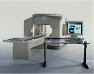
Manufactured by Esaote S.p.A.;
a low field open MRI scanner with permanent magnet for orthopedic use. The outstanding feature of this MRI system is a patient friendly design with 24 cm diameter, which allows the imaging of extremities and small body parts like shoulder MRI. The power consumption is around 1.3 kW and the needed minimum floor space is an area of 16 sq m.
At RSNA 2006 Hologic Inc. introduced a new dedicated extremity MRI scanner, the Opera. Manufactured by Esaote is the Opera a redesign of Esaote's 0.2 Tesla E-Scan XQ platform, which now enables complete imaging of all extremities, including hip and shoulder applications. 'Real-time positioning' reportedly speeds patient setup and reduces exam times.
Esaote North America and Hologic Inc are the U.S. distributors of this MRI device.
Device Information and Specification
CLINICAL APPLICATION
Dedicated extremity
CONFIGURATION
Extremity, shoulder (2), flex coil, knee dual phased array, ankle//foot dual phased array, hand//wrist dual phased array coil
PULSE SEQUENCES
SE, GE, IR, STIR, FSE, 3D CE, GE-STIR, 3D GE, ME, TME, HSE
IMAGING MODES
Single, multislice, volume study, fast scan, multi slab
TR
10 - 5000 msec
TE
6 - 110 msec
SINGLE SLICE
0.6 sec/image
MULTISLICE
0.6 sec/image
17 cm
2D: 2 mm - 10 mm;
3D: 0.6 mm - 10 mm
MEASURING MATRIX
256 x 256 maximum
PIXEL INTENSITY
4096 gray lvls, 256 lvls in 3D
MAGNET TYPE
Permanent
24 cm H, open
MAGNET WEIGHT
2250 kg, 4960 lbs
H*W*D
79 x 65 x 85 cm
POWER REQUIREMENTS
2,0 kW; 110/220 V single phase
STRENGTH
20 mT/m
5-GAUSS FRINGE FIELD, radial/axial
150 cm/130 cm
Passive
spacer

• View the DATABASE results for 'Opera (E-SCAN™ XQ)' (2).Open this link in a new window

 
Further Reading:
  News & More:
E-Scan, 510(k) Summary(.pdf)
Saturday, 15 May 2004   by www.accessdata.fda.gov    
MRI Resources 
Safety Training - IR - Journals - Abdominal Imaging - Examinations - MRI Reimbursement
 
Panorama 0.23T™InfoSheet: - Devices -
Intro, 
Types of Magnets, 
Overview, 
etc.MRI Resource Directory:
 - Devices -
 
www.medical.philips.com/main/products/mri/products/panoramafamily/panorama0.23t_rt/features/ From Philips Medical Systems;
the Panorama 0.23 T, providing a new design optimized for patient comfort, faster reconstruction time than before (300 images/second) and new gradient specifications. Philips' Panorama 0.23 T I/T supports MR-guided interventions, resulting in minimally invasive procedures, more targeted surgery, reduced recovery time and shorter hospital stays. Optional OptoGuide functionality enables real-time needle tracking. Philips' Panorama 0.23 TPanorama 0.2 R/T is the first and only open MRI system to enable radiation therapy planning using MR data sets. The Panorama also features the new and consistent Philips User Interface, an essential element of the Vequion clinical IT family of products and services.
Device Information and Specification
CLINICAL APPLICATION
Whole body
CONFIGURATION
Open MRI/C-arm
Head, head-neck, extremity M-L, neck, body/spine S-XL, shoulder, bilateral breast, wrist, TMJ, flex XS-S-M-L-XL-XXL
SYNCHRONIZATION
ECG/peripheral: Optional/optional, respiratory gating
PULSE SEQUENCES
SE, FE, IR, FFE, DEFFE, DESE, TSE, DETSE, Single shot SE, DRIVE, Balanced FFE, MRCP, Fluid Attenuated Inversion Recovery, Turbo FLAIR, IR-TSE, T1-STIR TSE, T2-STIR TSE, Diffusion Imaging, 3D SE, 3D FFE, MTC;; Angiography: CE-ANGIO, MRA 2D, 3D TOF
IMAGING MODES
Single, multislice, volume study, dynamic, SIMEX, multi chunk 3D, multiple stacks
TR
Min. 6.2 msec
TE
Min. 2.8 msec
SINGLE/MULTI SLICE
50 slices/sec
0.4 cm - 40 cm
1280 X 1024
MEASURING MATRIX
Up to 512 x 512
PIXEL INTENSITY
256 gray scale
MAGNET TYPE
Resistive/iron core
Open x 46 cm x infinite (side-first patient entry)
MAGNET WEIGHT
13110 kg
H*W*D
196 x 121 x 176 cm
POWER REQUIREMENTS
400/480 V
COOLING SYSTEM TYPE
Closed loop chilled water (chiller included)
N/A
STRENGTH
19 mT/m
5-GAUSS FRINGE FIELD
2.4 m / 3.7 m
Passive/active
spacer

• View the DATABASE results for 'Panorama 0.23T™' (2).Open this link in a new window

 
Further Reading:
  News & More:
Magnetic resonance imaging guided musculoskeletal interventions at 0.23T: Chapter 4. Materials and methods
2002
MRI Resources 
Mobile MRI Rental - Examinations - Pacemaker - Education - Abdominal Imaging - MRI Training Courses
 
Panorama 0.6TPanorama 0.2InfoSheet: - Devices -
Intro, 
Types of Magnets, 
Overview, 
etc.MRI Resource Directory:
 - Devices -
 
www.medical.philips.com/main/products/mri/products/panoramafamily/panorama0.23t_rt/features/ From Philips Medical Systems;
Panorama 0.6 T is the Philips Mid-Field Open MRI system. It is the most open MR scanner in the market, optimized for patient comfort and faster reconstruction time.
Device Information and Specification
CLINICAL APPLICATION
Whole body
CONFIGURATION
Open MRI/C-arm
Head, head-neck, extremity, neck, body/spine M-XL, shoulder, bilateral breast, wrist, TMJ, flex XS-S-M-L-XL-XXL
SYNCHRONIZATION
ECG/peripheral: Optional/yes, respiratory gating
PULSE SEQUENCES
SE, FE, IR, STIR, FFE, DEFFE, DESE, TSE, DETSE, Single shot SE, DRIVE, Balanced FFE, MRCP, Fluid Attenuated Inversion Recovery, Turbo FLAIR, IR-TSE, T1-STIR TSE, T2-STIR TSE, Diffusion Imaging, 3D SE, 3D FFE, Contrast Perfusion Analysis, MTC;; Angiography: CE-ANGIO, MRA 2D, 3D TOF
IMAGING MODES
Single, multislice, volume study, dynamic, SIMEX, multi chunk 3D, multiple stacks
TR
Min. 4.6 msec
TE
Min. 2.3 msec
SINGLE/MULTI SLICE
50 slices/sec
0.4 cm - 42 cm
1280 X 1024
MEASURING MATRIX
Up to 512 x 512
PIXEL INTENSITY
256 gray scale
MAGNET TYPE
Superconducting / iron core
Open x 47 cm x infinite (side-first patient entry)
MAGNET WEIGHT
38000 kg
H*W*D
254 x 244 x 325 cm
POWER REQUIREMENTS
400/480 V
COOLING SYSTEM TYPE
Liquid helium//air cool
0.00 L/hr helium
STRENGTH
20 mT/m
5-GAUSS FRINGE FIELD
2.4 m / 2.5 m
Passive/active
spacer

• View the DATABASE results for 'Panorama 0.6T™' (2).Open this link in a new window

MRI Resources 
Brain MRI - Shielding - Libraries - Jobs - Pediatric and Fetal MRI - Artifacts
 
Panorama 1.0T™InfoSheet: - Devices -
Intro, 
Types of Magnets, 
Overview, 
etc.MRI Resource Directory:
 - Devices -
 
www.medical.philips.com/main/products/mri/products/panoramafamily/panorama1.0t_rt/features/ From Philips Medical Systems;
this active shielded member of the Panorama product line combines the advantages of one 1.0 T system's with the possibilities of an open MRI system. The open design helps ease anxiety for claustrophobic patients and increased patient comfort whereby the field strength provides spectacular image quality and fast patient throughput.
Device Information and Specification
CLINICAL APPLICATION
Whole body
CONFIGURATION
Vertical Field open MRI post design
Vertically opposed solenoids, head, head-neck, extremity, neck, body/spine M-XL, shoulder, bilateral breast, wrist, TMJ, flex XS-S-M-L-XL-XXL
SYNCHRONIZATION
ECG/peripheral: Optional/yes, respiratory gating
PULSE SEQUENCES
SE, FE, IR, STIR, FFE, DEFFE, DESE, TSE, DETSE, Single shot SE, DRIVE, Balanced FFE, MRCP, FLAIR, Turbo FLAIR, IR-TSE, T1-STIR TSE, T2-STIR TSE, Diffusion Imaging, 3D SE, 3D FFE, Contrast Perfusion Analysis, MTC;; Angiography: CE-ANGIO, MRA 2D, 3D TOF
IMAGING MODES
Single, multislice, volume study, dynamic, SIMEX, multi chunk 3D, multiple stacks
TR
Min. 4.6 msec
TE
Min. 2.3 msec
SINGLE/MULTI SLICE
50 slices/sec
0.4 cm - 42 cm
1280 X 1024
MEASURING MATRIX
Up to 512 x 512
PIXEL INTENSITY
256 gray scale
Open x 47 cm x infinite (side-first patient entry)
MAGNET WEIGHT
H*W*D
POWER REQUIREMENTS
400/480 V
COOLING SYSTEM TYPE
Liquid helium
STRENGTH
20 mT/m
5-GAUSS FRINGE FIELD
Passive/active
spacer

• View the DATABASE results for 'Panorama 1.0T™' (2).Open this link in a new window

MRI Resources 
Portals - Non-English - Corporations - DICOM - MRI Centers - IR
 
previous      36 - 40 (of 55)     next
Result Pages : [1 2 3 4 5 6 7 8 9 10 11]
 Random Page
 
Share This Page
FacebookTwitterLinkedIn

MR-TIP    
Community   
User
Pass
Forgot your UserID/Password ?    


New acceleration techniques will :
reduce scan times 
cause artifacts 
increase expenses 
be useful if you have a lot of experience 
doesn't do much 
never heard of 

Look
      Ups


MR-TIP.com uses cookies! By browsing MR-TIP.com, you agree to our use of cookies.

Magnetic Resonance - Technology Information Portal
Member of SoftWays' Medical Imaging Group - MR-TIP • Radiology-TIP • Medical-Ultrasound-Imaging • 
Copyright © 2003 - 2024 SoftWays. All rights reserved. [ 16 April 2024]
Terms of Use | Privacy Policy | Advertising
 [last update: 2024-02-26 03:41:00]