Magnetic Resonance - Technology Information Portal Welcome to MRI Technology
Info
  Sheets

Out-
      side


 'Boil%20off%20Rate' 
SEARCH FOR    
 
  2 3 5 A B C D E F G H I J K L M N O P Q R S T U V W X Y Z
Result : Searchterm 'Boil off Rate' found in 1 term [] and 11 definitions [], (+ 3 Boolean[] results
1 - 5 (of 15)     next
Result Pages : [1]  [2 3]
MRI Resources 
MRA - Collections - Examinations - DICOM - Services and Supplies - Breast Implant
 
Boil off Rate
 
Rate of cryogen (liquid helium) evaporation in superconducting magnets, usually measured in liters of liquid per hour. The boil off rate of cryogen increases during ramping of the magnet and with eddy currents induced in the cryoshields by pulsed field gradients. In calculating cryogen consumption additional transfer and filling losses have to be considered.

See also Cryoshielding and Helium.
spacer
 
• Share the entry 'Boil off Rate':  Facebook  Twitter  LinkedIn  
 
Further Reading:
  Basics:
Cryogenic Liquids and their Hazards
   by www.ccohs.ca    
Zero Boil Off Cryostats
Friday, 23 September 2011   by www.cryogenicsociety.or    
  News & More:
A hot time for cold superconductors
Tuesday, 9 December 2003   by www.brightsurf.com    
MRI Resources 
Health - Universities - Safety pool - Brain MRI - Spine MRI - Mobile MRI
 
Aurora® 1.5T Dedicated Breast MRI SystemInfoSheet: - Devices -
Intro, 
Types of Magnets, 
Overview, 
etc.
 
www.auroramri.com/mri/product/ From Aurora Imaging Technology, Inc.;
The Aurora® 1.5T Dedicated Breast MRI System with Bilateral SpiralRODEO™ is the first and only FDA approved MRI device designed specifically for breast imaging. The Aurora System, which is already in clinical use at a growing number of leading breast care centers in the US, Europe, got in December 2006 also the approval from the State Food and Drug Administration of the People's Republic of China (SFDA).
'Some of the proprietary and distinguishing features of the Aurora System include: 1) an ellipsoid magnetic shim that provides coverage of both breasts, the chest wall and bilateral axillary lymph nodes; 2) a precision gradient coil with the high linearity required for high resolution spiral reconstruction;; 3) a patient-handling table that provides patient comfort and procedural utility; 4) a fully integrated Interventional System for MRI guided biopsy and localization; and 5) the user-friendly AuroraCAD™ computer-aided image display system designed to improve the accuracy and efficiency of diagnostic interpretations.'
Device Information and Specification
CLINICAL APPLICATION
CONFIGURATION
Short bore compact
IMAGING MODES
Bi-lateral RODEO fat suppression, high resolution Rotating Delivery of Excitation Offresonance Spiral, integrated targeting SW compatible with major MR guided intervention equipment
TR
10 ms for gradient echo and less than 2,500 ms for T2 weighted spin echo
TE
From 5 ms for RODEO Plus to over 80 ms, 120 ms for T2 sequences
SINGLE/MULTI SLICE
Around 0.02 sec for a 256x256 image, 12.4 sec for a 512 x 512 x 32 multislice set
FOV
20 - 36 cm, max. elliptical 36 x 44 cm
MEASURING MATRIX
512 x 512
BORE DIAMETER
or W x H
64 cm diameter (gantry)
MAGNET WEIGHT
8,500 lbs
H*W*D
240 x 188 x 163 cm
POWER REQUIREMENTS
150A/120V-208Y/3 Phase//60 Hz/5 Wire
COOLING SYSTEM
Helium for magnet, distilled/de-ionized water for coil;
spacer

• View the DATABASE results for 'Aurora® 1.5T Dedicated Breast MRI System' (2).Open this link in a new window


• View the NEWS results for 'Aurora® 1.5T Dedicated Breast MRI System' (3).Open this link in a new window.
 
Further Reading:
  News & More:
Aurora Imaging Technology Announces Approval in China
   by salesandmarketingnetwork.com    
Aurora Imaging Technology, Inc. Release: Results of a Multi-Center Trial Demonstrates Superior Diagnostic Performance of the Aurora® 1.5T Dedicated Breast MRI System Over Whole-Body Breast MRI
Monday, 1 October 2012   by www.biospace.com    
MRI Resources 
General - Open Directory Project - Image Quality - Chemistry - Used and Refurbished MRI Equipment - Stimulator pool
 
Biograph mMR
 
www.healthcare.siemens.com/magnetic-resonance-imaging/mr-pet-scanner/biograph-mmr 
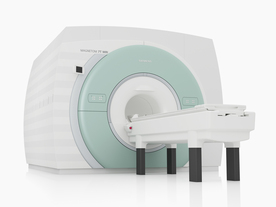
FDA cleared and CE Mark 2011.
The Biograph mMR has a fully-integrated design for simultaneous PET/MRI imaging. The dedicated hardware includes solid-state, avalanche photodiode PET detector and adapted, PET-compatible MR coils.
The possibility of truly simultaneous operation allows the acquisition of several magnetic resonance imaging (MRI) sequences during the positron emission tomography (PET) scan, without increasing the examination time.
See also Hybrid Imaging.
Device Information and Specification
CLINICAL APPLICATION
Whole Body
CONFIGURATION
Simultaneous PET/MRI
3 Tesla
Full range of MRI coils
CHANNELS
up to 32
PET ACQUISITION MODES
3D
MRI ACQUISITION MODES
2D/3D, spectroscopy;; iPAT, GRAPPA (k-space), noncontrast angiography, radial motion compensation
PET EFFECTIVE AXIAL FOV
26 cm (typical overlap 23%)
MRI FOV
A-P 45, R-L 50, H-F 50 cm
PET RING DIAMETER
65.6 cm
PATIENT SCAN RANGE
199 cm
HORIZONTAL SPEED
200 mmsec
TABLE CAPACITY
200 kg
PET DETECTOR
Solid state, 4032 avalanche photo diodes
DETECTOR SCINTILLATION MATERIAL
LSO, 28672 crystals
CRYSTAL SIZE
4 x 4 x 20 mm
MAGNET WEIGHT
9000 kg
DIMENSION H*W*D (gantry included)
335 x 230 x 242 cm (finshed covers)
5-GAUSS FRINGE FIELD
2.6 m / 4.6 m
CRYOGEN USE
Zero boil off rate, approx. 10 years
COOLING SYSTEM
PET system: water; MRI system: water
up to 200 T/m/s
MAX. AMPLITUDE
45 mT/m
Aautomatic, patient specific shim; active shim 3 linear and 5 non-linear channels (seond order)
POWER REQUIREMENTS
380 / 400 / 420 / 440 / 460 / 480 V, 3-phase + ground; Total system 110kW
spacer
 
Further Reading:
  Basics:
Performance Measurements of the Siemens mMR Integrated Whole-Body PET/MR Scanner
Friday, 11 November 2011   by jnm.snmjournals.org    
Global Trends in Hybrid Imaging
Monday, 1 November 2010   by pubs.rsna.org    
  News & More:
Positron Emission Tomographic Imaging in Stroke
Monday, 28 December 2015   by www.ncbi.nlm.nih.gov    
A world's first in imaging – integrated whole-body molecular MR system available for clinical use testing
Thursday, 18 November 2010   by www.siemens.com    
FDA Clears New System to Perform Simultaneous PET, MRI Scans
Friday, 10 June 2011   by www.prnewswire.com    
Frost & Sullivan Award Underlines Siemens Healthcare's Contribution to New Product Innovation in the North American Medical Imaging Market
Wednesday, 13 July 2011   by multivu.prnewswire.com    
MRI Resources 
Universities - Health - Quality Advice - Claustrophobia - Pediatric and Fetal MRI - Calculation
 
Cryostat
 
Cryostats are devices for maintaining a constant low temperature (as by means of liquid helium used in MRI). They require vacuum chambers with thermal isolation.

See also Dewar, Cryoshielding and Boil off Rate.
spacer

• View the DATABASE results for 'Cryostat' (6).Open this link in a new window

 
Further Reading:
  Basics:
Superconducting Magnets
   by mri-q.com    
Zero Boil Off Cryostats
Friday, 23 September 2011   by www.cryogenicsociety.or    
MRI Resources 
Libraries - Journals - Mass Spectrometry - PACS - MRA - Breast Implant
 
MAGNETOM 7T
 
www.healthcare.siemens.com/magnetic-resonance-imaging/7t-mri-scanner/magnetom-7t From Siemens Medical Systems;
The MAGNETOM 7T is designed as an open research platform. 7T MRI provides anatomical detail at the submillimiter scale, enhanced contrast mechanisms, outstanding spectroscopy performance, ultra-high resolution functional imaging (fMRI), multinuclear whole-body MRI and functional information.
This ultra high field (UHF) MRI device is a research system and not cleared, approved or licensed in any jurisdiction for patient examinations.
Device Information and Specification
CLINICAL APPLICATION
Whole body
CONFIGURATION
Compact
7 Tesla
High-performance, ultra high field coils available. Integration and support for coil developments.
CHANNELS (min. / max. configuration)
32, optional 8 channels TX array
Chemical shift imaging, single voxel spectroscopy, multinuclear imaging optional
IMAGING TECHNIQUES
iPAT, mSENSE and GRAPPA (image, k-space), noncontrast angiography, plaque imaging, radial motion compensation
FOV
40 x 40 x 30 cm³ less than 8% nonlinearity
BORE DIAMETER
or W x H
60 cm
TABLE CAPACITY
200 kg
MAGNET WEIGHT (gantry included)
35017 kg
DIMENSION H*W*D (gantry included)
320 x 240 x 317,5 cm
5-GAUSS FRINGE FIELD
7.9 m / 5.6 m
CRYOGEN USE
Zero boil off rate
COOLING SYSTEM
Water
up to 200 T/m/s
MAX. AMPLITUDE
up to 70 mT/m
Up to 3rd order shim coils, user configurable B0 shim ? B0 maps and ROI definition
POWER REQUIREMENTS
2000 Volts, 650A
spacer
 
Further Reading:
  Basics:
MAGNETOM 7T Product Brochure
   by www.healthcare.siemens.com    
  News & More:
Ultra-high-field MRI may allow earlier diagnosis of Parkinson's disease
Wednesday, 5 March 2014   by www.sciencedaily.com    
Feasibility of Using Ultra-High Field (7 T) MRI for Clinical Surgical Targeting
Thursday, 17 May 2012   by www.plosone.org    
Ultrahigh-Field MRI May Detect Additional Pathology in EAE
Sunday, 20 October 2013   by www.msdiscovery.org    
MRI Resources 
Musculoskeletal and Joint MRI - Spectroscopy pool - Software - Equipment - Portals - Brain MRI
 
     1 - 5 (of 15)     next
Result Pages : [1]  [2 3]
 Random Page
 
Share This Page
FacebookTwitterLinkedIn

MR-TIP    
Community   
User
Pass
Forgot your UserID/Password ?    


How AI will impact MRI :
only diagnostics 
saving time 
reducing cost 
makes planning obsolete 
reduce human knowledge 
not at all 

Look
      Ups


MR-TIP.com uses cookies! By browsing MR-TIP.com, you agree to our use of cookies.

Magnetic Resonance - Technology Information Portal
Member of SoftWays' Medical Imaging Group - MR-TIP • Radiology-TIP • Medical-Ultrasound-Imaging • 
Copyright © 2003 - 2024 SoftWays. All rights reserved. [ 20 April 2024]
Terms of Use | Privacy Policy | Advertising
 [last update: 2024-02-26 03:41:00]