Magnetic Resonance - Technology Information Portal Welcome to MRI Technology
Info
  Sheets

Out-
      side


 'Angiography' 
SEARCH FOR    
 
  2 3 5 A B C D E F G H I J K L M N O P Q R S T U V W X Y Z
Result : Searchterm 'Angiography' found in 14 terms [] and 98 definitions []
previous     41 - 45 (of 112)     next
Result Pages : [1 2 3]  [4 5 6 7 8 9 10 11 12 13 14 15 16 17 18 19 20 ... ]
Searchterm 'Angiography' was also found in the following services: 
spacer
News  (19)  Resources  (26)  Forum  (6)  
 
Excelart AG™ with PianissimoInfoSheet: - Devices -
Intro, 
Types of Magnets, 
Overview, 
etc.MRI Resource Directory:
 - Devices -
 
http://www.medical.toshiba.com/clinical/radiology/15texcelart.htm From Toshiba America Medical Systems Inc.;
the EXCELART is a superconducting whole body MRI system with a short wide-bore magnet, operating at 1.5 T. It features powerful high-speed gradients with a revolutionary gradient acoustic noise reduction system: Pianissimo. The dramatic reduction of gradient acoustic noise by Pianissimo greatly enhances patient comfort during exams. The standard array platform and a wide range of array coils ensure excellent images. A powerful 64-bit RISC-based computer system and newly developed array processor realize high productivity.
Device Information and Specification
CLINICAL APPLICATION
Whole body
CONFIGURATION
Cylindrical Wide Short Bore
Opt. (WIP) Single and Multi Voxel
SYNCHRONIZATION
ECG: standard; peripheral: optional, respiratory gating
PULSE SEQUENCES
SE, FE, IR, FastSE, FastIR, FastFLAIR, Fast STIR, FastFE, FASE, Hybrid EPI, Multi Shot EPI; Angiography: 2D(gate/non-gate)/3D TOF, SORS-STC
IMAGING MODES
Single, multislice, volume study
TR
3.5-30000 msec
TE
8 msec min. SE; 1.2 msec min. FE
SINGLE/MULTI SLICE
less than 0.015 (256x256)
FOV
2 cm
1.0 min. 2-DFT: 0.2 min. 3-DFT
Up to 1024
MEASURING MATRIX
32-1024, phase;; 64-1024, freq.
PIXEL INTENSITY
256 gray levels
BORE DIAMETER
or W x H
65.5 cm, patient aperture
MAGNET WEIGHT
4050 kg (bare magnet incl. L-He)
H*W*D
235 x 219 x 199 cm
COOLING SYSTEM TYPE
Closed-loop water-cooled
CRYOGEN USE
Liquid helium: approx. less than 0.05 L/hr
STRENGTH
25 mT/m
5-GAUSS FRINGE FIELD
2.5 m / 4.0 m
Passive, active, auto-active
spacer
Searchterm 'Angiography' was also found in the following services: 
spacer
Radiology  (34) Open this link in a new windowUltrasound  (9) Open this link in a new window
Excelart XG™ with PianissimoInfoSheet: - Devices -
Intro, 
Types of Magnets, 
Overview, 
etc.MRI Resource Directory:
 - Devices -
 
http://www.medical.toshiba.com/clinical/radiology/15texcelart.htm From Toshiba America Medical Systems Inc.;
the EXCELART is a superconducting whole body MRI system with a short wide-bore magnet, operating at 1.5 T. It features powerful high-speed gradients with a revolutionary gradient acoustic noise reduction system: Pianissimo. The dramatic reduction of gradient acoustic noise by Pianissimo greatly enhances patient comfort during exams. The standard array platform and a wide range of array coils ensure excellent images. A powerful 64-bit RISC-based computer system and newly developed array processor realize high productivity.
Device Information and Specification
CLINICAL APPLICATION
Whole body
CONFIGURATION
Cylindrical Wide Short Bore
Optional (WIP)
SYNCHRONIZATION
ECG/peripheral, respiratory gating
PULSE SEQUENCES
SE, FE, IR, FastSE, FastIR, FastFLAIR, Fast STIR, FastFE, FASE, Hybrid EPI, Multi Shot EPI; Angiography: 2D(gate/non-gate)/3D TOF, SORS-STC
IMAGING MODES
Single, multislice, volume study
TR
2.6-30000 msec
TE
8 msec min. SE; 0.9 msec min. FE
SINGLE/MULTI SLICE
less than 0.011 (256x256)
FOV
2 cm
1.0 min. 2-DFT: 0.2 min. 3-DFT
Up to 1024
MEASURING MATRIX
32-1024, phase;; 64-1024, freq.
PIXEL INTENSITY
256 gray levels
BORE DIAMETER
or W x H
65.5 cm, patient aperture
MAGNET WEIGHT
4050 kg (bare magnet incl. L-He)
H*W*D
235 x 219 x 199 cm
POWER REQUIREMENTS
380/400/415/440/480 V
COOLING SYSTEM TYPE
Closed-loop water-cooled
CRYOGEN USE
Liquid helium: approx. less than 0.05 L/hr
STRENGTH
25 mT/m
5-GAUSS FRINGE FIELD
2.5 m / 4.0 m
Passive, active, auto-active
spacer

• View the DATABASE results for 'Excelart XG™ with Pianissimo' (2).Open this link in a new window

 
Further Reading:
  News & More:
Angio, cardiac imaging top list for MR and CT
1999   by www.diagnosticimaging.com    
MRI Resources 
IR - Shielding - - Implant and Prosthesis - Research Labs - Crystallography
 
FLEXART™InfoSheet: - Devices -
Intro, 
Types of Magnets, 
Overview, 
etc.MRI Resource Directory:
 - Devices -
 
From Toshiba America Medical Systems Inc.; www3.toshiba.co.jp/medical/products/mr/flex/index.htm 
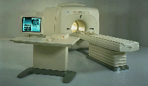
FLEXART™ series is a 0.5 T superconducting MRI system that has been designed to meet the expanding role of MRI in today's clinical environment. The system utilizes innovative technologies such as digital RF, high speed actively shielded gradients and optimized RF coils which support a wide range of MRI developments.
Device Information and Specification
CLINICAL APPLICATION
Whole body
CONFIGURATION
Compact
Quadrature, solenoid and multi-channel configurations
SYNCHRONIZATION
ECG/peripheral optional, respiratory gating
PULSE SEQUENCES
SE, FE, IR, FastSE, FastIR, FastFLAIR, Fast STIR, FastFE, FASE, Hybrid EPI, Multi Shot EPI; Angiography: 2D(gate/non-gate)/3D TOF, SORS-STC
IMAGING MODES
Single, multislice, volume study
POWER REQUIREMENTS
380/400/415/440/480 V
COOLING SYSTEM TYPE
Closed-loop water-cooled
STRENGTH
25 mT/m
Passive, active
spacer

• View the DATABASE results for 'FLEXART™' (2).Open this link in a new window

Searchterm 'Angiography' was also found in the following services: 
spacer
News  (19)  Resources  (26)  Forum  (6)  
 
FORTE 3.0T™InfoSheet: - Devices -
Intro, 
Types of Magnets, 
Overview, 
etc.MRI Resource Directory:
 - Devices -
 
www.isoltech.co.kr/english/product/30t.htm From ISOL Technology
'Ultra high field MR system, it's right close to you. FORTE 3.0T is the new standard for the future ultra high field MR system. If you are pushing the limits of your existing clinical MR scanner, the FORTE will surely take you to the next level of diagnostic imaging. FORTE is the core leader of the medical technology in the 21st century. Proving effects of fMRI that cannot be measured with MRI less than 2.0T.'
Device Information and Specification
CLINICAL APPLICATION
Whole body
CONFIGURATION
Short bore compact
SYNCHRONIZATION
ECG/peripheral: Optional/yes, external trigger, respiratory gating
PULSE SEQUENCES
Spin echo, Gradient echo, Fast spin echo, Inversion recovery, 2D/3D Fast gradient echo sequences FLAIR/STIR, 2D/3D TOF
IMAGING MODES
2D/3D, T1, T2 and Diffusion//Perfusion imaging, MR Angiography package, Advanced EPI package, Multi-nuclei MR Spectroscopy package
FOV
40 cm
128 x 128, 256 x 256, 512 x 512, 1024 x 1024
BORE DIAMETER
or W x H
61 cm without body coil
MAGNET WEIGHT
12000 kg
H*W*D
260 x 220 x 235 cm
COOLING SYSTEM TYPE
Water-cooled coil and air-cooled amplifier
CRYOGEN USE
0.15 L/hr helium
STRENGTH
38 mT/m
5-GAUSS FRINGE FIELD
3.3 m / 5.2 m
Passive and active
spacer

• View the DATABASE results for 'FORTE 3.0T™' (2).Open this link in a new window

Searchterm 'Angiography' was also found in the following services: 
spacer
Radiology  (34) Open this link in a new windowUltrasound  (9) Open this link in a new window
Fast Imaging with Steady State PrecessionInfoSheet: - Sequences - 
Intro, 
Overview, 
Types of, 
etc.
 
(FISP) A fast imaging sequence, which attempts to combine the signals observed separately in the FADE sequence, generally sensitive about magnetic susceptibility artifacts and imperfections in the gradient waveforms. Confusingly now often used to refer to a refocused FLASH type sequence.
This sequence is very similar to FLASH, except that the spoiler pulse is eliminated. As a result, any transverse magnetization still present at the time of the next RF pulse is incorporated into the steady state. FISP uses a RF pulse that alternates in sign. Because there is still some remaining transverse magnetization at the time of the RF pulse, a RF pulse of a degree flips the spins less than a degree from the longitudinal axis. With small flip angles, very little longitudinal magnetization is lost and the image contrast becomes almost independent of T1. Using a very short TE (with TR 20-50 ms, flip angle 30-45°) eliminates T2* effects, so that the images become proton density weighted. As the flip angle is increased, the contrast becomes increasingly dependent on T1 and T2*. It is in the domain of large flip angles and short TR that FISP exhibits vastly different contrast to FLASH type sequences. Used for T1 orthopedic imaging, 3D MPR, cardiography and angiography.
spacer

• View the DATABASE results for 'Fast Imaging with Steady State Precession' (5).Open this link in a new window

 
Further Reading:
  Basics:
MRI techniques improve pulmonary embolism detection
Monday, 19 March 2012   by medicalxpress.com    
MRI Resources 
Quality Advice - Homepages - Movies - Services and Supplies - Functional MRI - Knee MRI
 
previous      41 - 45 (of 112)     next
Result Pages : [1 2 3]  [4 5 6 7 8 9 10 11 12 13 14 15 16 17 18 19 20 ... ]
 Random Page
 
Share This Page
FacebookTwitterLinkedIn

MR-TIP    
Community   
User
Pass
Forgot your UserID/Password ?    


Personalized protocols (age, gender, body habitus, etc.) lead to :
more automated planning 
improved patient comfort 
better diagnostics 
optimized image quality 
nothing 

Look
      Ups


MR-TIP.com uses cookies! By browsing MR-TIP.com, you agree to our use of cookies.

Magnetic Resonance - Technology Information Portal
Member of SoftWays' Medical Imaging Group - MR-TIP • Radiology-TIP • Medical-Ultrasound-Imaging • 
Copyright © 2003 - 2024 SoftWays. All rights reserved. [ 19 April 2024]
Terms of Use | Privacy Policy | Advertising
 [last update: 2024-02-26 03:41:00]