Magnetic Resonance - Technology Information Portal Welcome to MRI Technology
Info
  Sheets

Out-
      side


 '5 Gauss Fringe Field' 
SEARCH FOR    
 
  2 3 5 A B C D E F G H I J K L M N O P Q R S T U V W X Y Z
Result : Searchterm '5 Gauss Fringe Field' found in 1 term [] and 7 definitions [], (+ 14 Boolean[] results
previous     11 - 15 (of 22)     next
Result Pages : [1]  [2]  [3 4 5]
MRI Resources 
MRI Technician and Technologist Career - Case Studies - Education pool - Resources - Pregnancy - Manufacturers
 
C-SCAN™InfoSheet: - Devices -
Intro, 
Types of Magnets, 
Overview, 
etc.MRI Resource Directory:
 - Devices -
 
www.gehealthcare.com/usen/mr/cscan/index.html Manufactured by Esaote S.p.A.; compact in-office MRI system, fits in a 90 ft2 (8.4 m2) space and requires no shielding or special power. This low field MRI magnet is optimized for orthopedic use and imaging of the extremities.
The C-SCAN™ is developed from the ARTOSCAN™ - M with a new computer platform, and is also known as Artoscan C.
Esaote North America and Hologic Inc. are the U.S. distributors of this MRI device.
Device Information and Specification
CLINICAL APPLICATION
Dedicated extremity
CONFIGURATION
Closed
Linear and dual phased array, knee, ankle, wrist (6 total) coils
PULSE SEQUENCES
SE, GE, IR, STIR, FSE, 3D CE, GE-STIR, 3D GE, ME, TME, HSE
IMAGING MODES
Single, multislice, volume study, fast scan, multi slab
TR
12 - 5,000 msec
TE
5 - 220 msec
SINGLE SLICE
0.8 sec/image
MULTISLICE
0.8 sec/image
FOV
10 - 20 cm
2D: 2 mm - 10 mm;
3D: 0.6 mm - 10 mm
512 x 512
MEASURING MATRIX
256 x 256 maximum
PIXEL INTENSITY
4,096 gray lvls, 256 lvls in 3D
MAGNET TYPE
Permanent
BORE DIAMETER
or W x H
33.6 x 16 cm
MAGNET WEIGHT
960 kg
H*W*D
124 x 76 x 60 cm
POWER REQUIREMENTS
100/110/200/220/230/240
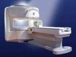
STRENGTH
10 mT/m
5-GAUSS FRINGE FIELD, radial/axial
28 cm/60 cm
Passive
spacer
MRI Resources 
Cochlear Implant - Journals - Blood Flow Imaging - Safety Training - Movies - Contrast Agents
 
Signa Ovation™InfoSheet: - Devices -
Intro, 
Types of Magnets, 
Overview, 
etc.MRI Resource Directory:
 - Devices -
 
www.gehealthcare.com/usen/mr/ovation/index.html From GE Healthcare;
the Signa Ovation™ is a patient-friendly open MRI scanner designed not only to handle a typical patient mix, but to accommodate larger patients, patients who are claustrophobic, and others who have difficulty tolerating the close quarters of conventional MR machines.
Device Information and Specification
CLINICAL APPLICATION
Whole body
CONFIGURATION
Integrated transmit and receive body coil; head coil, 4 channel neurovascular array, 8 channel CTL array, 2 channel shoulder array, 3 channel large extremity array, 3 channel small extremity array, 4 channel foot array, 3 channel wrist array
SYNCHRONIZATION
Standard cardiac gating, ECG/peripheral, respiratory gating
PULSE SEQUENCES
Standard: SE, IR, 2D/3D GRE and SPGR, 2D/3D TOF, 2D/3D FSE, 2D/3D FGRE and FSPGR, SSFP, FLAIR, EPI, optional: 2D/3D Fiesta, true chem sat, fat/water separation, single shot diffusion EPI, line scan diffusion
IMAGING MODES
Localizer, single slice, multislice, volume, fast, POMP, multi slab, cine, slice and frequency zip, extended dynamic range, tailored RF
TR
1.3 to 12000 msec in increments of 1 msec
TE
0.4 to 2000 msec in increments of 1 msec
SINGLE/MULTI SLICE
Simultaneous scan and reconstruction;; 80 images/second reconstruction
3cm to 40 cm continuous
2D: 1.4mm - 20mm 3D: 0.2mm - 20mm
1280 x 1024
MEASURING MATRIX
128x512 steps 32 phase//freq.
PIXEL INTENSITY
256 gray levels
0.08 mm; 0.02 mm optional
MAGNET TYPE
Permanent
175 x 85 x 447 cm
MAGNET WEIGHT
19200 kg
H*W*D
530 x 175 x 250 cm
POWER REQUIREMENTS
200 - 480, 3-phase
GRADIENT COOLING
None
MAX. GRADIENT AMPLITUDE
19 mT/m
5-GAUSS FRINGE FIELD
2.5 m/2.5 m
Computerized passive shimming during magnet setup, autoshim per series with automatic table motion to magnet isocenter for each prescription
spacer

• View the DATABASE results for 'Signa Ovation™' (2).Open this link in a new window

MRI Resources 
RIS - Pacemaker - MRI Physics - Colonography - Homepages - Pediatric and Fetal MRI
 
MSK-Extreme™InfoSheet: - Devices -
Intro, 
Types of Magnets, 
Overview, 
etc.MRI Resource Directory:
 - Devices -
 
www.onicorp.com/ From ONI Medical Systems, Inc.;
MSK-Extreme™MRI system is a dedicated high field extremity imaging device, designed to provide orthopedic surgeons and other physicians with detailed diagnostic images of the foot, ankle, knee, hand, wrist and elbow, all with the clinical confidence and advantages derived from high field, whole body MRI units. The light weight (less than 650 kg) of the OrthOne System performs rapid patient studies, is easy to operate, has a patient friendly open environment and can be installed in a practice office or hospital, all at a cost similar to a low field extremity machine.
New features include a more powerful operating system that offers increased scan speed as well as a 160-mm knee coil with higher signal to noise ratio, and the option of a CD burner.
Device Information and Specification
CLINICAL APPLICATION
Dedicated extremity imaging
CONFIGURATION
16 cm knee, 18 cm lower extremity;; 12.3 cm upper extremity, additional high resolution v-SPEC Coils: 80 mm, 100 mm, or 145 mm.
SYNCHRONIZATION
No
PULSE SEQUENCES
SE, FSE, GE2D, GE3D, Inversion recovery (IR), Driven Equilibrium, Fat Saturation (FS), STIR, MT, PD, Flow Compensation (FC), RF spoiling, MTE, No Phase Wrap (NPW)
IMAGING MODES
Scout, single, multislice, volume
TR
10-10,000ms; 1ms steps
TE
5-150ms; 1 ms steps
SINGLE/MULTI SLICE
2D less than 200 msec/image
4cm-16cm
2D: 2mm-10mm/.1mm incr.
Up to 1,000x1,000
MEASURING MATRIX
X/Y: 64-512; 2 pixel steps
PIXEL INTENSITY
4,096 grey lvls; 256 lvls in 3D
28cm ID x 50cm L
MAGNET WEIGHT
635 kg
H*W*D
146 x 69 x 84 cm
POWER REQUIREMENTS
115VAC, 1phase, 20A; 208VAC, 3 phase, 30A
COOLING SYSTEM TYPE
LHe with 2 stage cold head
Negligible
STRENGTH
15 mT/m
5-GAUSS FRINGE FIELD
1.25m radial x 1.8m axial
Passive
spacer
 
Further Reading:
  Basics:
MSK Extreme Brochure(.pdf)
   by www.nova-logic.ch    
MSK Extreme Specifications(.pdf)
   by www.nova-logic.ch    
MRI Resources 
MRI Reimbursement - Safety Products - Raman Spectroscopy - Patient Information - Cardiovascular Imaging - Distributors
 
Intera Achieva 1.5T™InfoSheet: - Devices -
Intro, 
Types of Magnets, 
Overview, 
etc.MRI Resource Directory:
 - Devices -
 
www.medical.philips.com/main/products/mri/products/intera_achieva/ From Philips Medical Systems;
The clinical capabilities of MR will further expand. Inside and out, the Achieva is a friendly, open system designed for optimal patient comfort and maximized workflow with high functionality. The Achieva 1.5T can be upgraded to Achieva I/T, with three configurations optimized for MR guided interventions and therapy:
Achieva I/T
Achieva I/T Neurosurgery
Achieva I/T Cardiovascular (or XMR - combining an Achieva 1.5T CV system and an X-Ray system)
Device Information and Specification
CLINICAL APPLICATION
Whole body
CONFIGURATION
Short bore compact
Standard: Head, body, C1, C3; Optional: Small joint, flex-E, flex-R, endocavitary (L and S), dual TMJ, knee, neck, T/L spine, breast; optional phased array: Spine, pediatric, 3rd party connector; Optional SENSEâ„¢ coils for all applications
Optional
SYNCHRONIZATION
ECG/peripheral: Optional/yes, respiratory gating
PULSE SEQUENCES
SE, Modified-SE, IR (T1, T2, PD), STIR, FLAIR, SPIR, FFE, T1-FFE, T2-FFE, Balanced FFE, TFE, Balanced TFE, Dynamic, Keyhole, 3D, Multi Chunk 3D, Multi Stack 3D, K Space Shutter, MTC, TSE, Dual IR, DRIVE, EPI, Cine, 2DMSS, DAVE, Mixed Mode; Angiography: Inflow MRA, TONE, PCA, CE MRA
IMAGING MODES
Single Slice 2D , Multi Single Slice 2D, Multi Slice 2D, 3D, Multi Chunk 3D, Multi Stack 3D
FOV
Over 40 cm
0.05 mm
128 x 128, 256 x 256,512 x 512,1024 x 1024 (64 for Bold img)
MEASURING MATRIX
Variable in 1% increments
PIXEL INTENSITY
Lum.: 120 cd/m2; contrast: 150:1
Variable (op. param. depend.)
60 x 60 cm
POWER REQUIREMENTS
380/400 V
CRYOGEN USE
0.03 L/hr helium
STRENGTH
up to 66 mT/m
5-GAUSS FRINGE FIELD
2.4 m / 3.8 m
Passive and dynamic
spacer

• View the DATABASE results for 'Intera Achieva 1.5T™' (2).Open this link in a new window

MRI Resources 
Service and Support - PACS - Online Books - Diffusion Weighted Imaging - Resources - Anatomy
 
Excelart XG™ with PianissimoInfoSheet: - Devices -
Intro, 
Types of Magnets, 
Overview, 
etc.MRI Resource Directory:
 - Devices -
 
http://www.medical.toshiba.com/clinical/radiology/15texcelart.htm From Toshiba America Medical Systems Inc.;
the EXCELART is a superconducting whole body MRI system with a short wide-bore magnet, operating at 1.5 T. It features powerful high-speed gradients with a revolutionary gradient acoustic noise reduction system: Pianissimo. The dramatic reduction of gradient acoustic noise by Pianissimo greatly enhances patient comfort during exams. The standard array platform and a wide range of array coils ensure excellent images. A powerful 64-bit RISC-based computer system and newly developed array processor realize high productivity.
Device Information and Specification
CLINICAL APPLICATION
Whole body
CONFIGURATION
Cylindrical Wide Short Bore
Optional (WIP)
SYNCHRONIZATION
ECG/peripheral, respiratory gating
PULSE SEQUENCES
SE, FE, IR, FastSE, FastIR, FastFLAIR, Fast STIR, FastFE, FASE, Hybrid EPI, Multi Shot EPI; Angiography: 2D(gate/non-gate)/3D TOF, SORS-STC
IMAGING MODES
Single, multislice, volume study
TR
2.6-30000 msec
TE
8 msec min. SE; 0.9 msec min. FE
SINGLE/MULTI SLICE
less than 0.011 (256x256)
FOV
2 cm
1.0 min. 2-DFT: 0.2 min. 3-DFT
Up to 1024
MEASURING MATRIX
32-1024, phase;; 64-1024, freq.
PIXEL INTENSITY
256 gray levels
BORE DIAMETER
or W x H
65.5 cm, patient aperture
MAGNET WEIGHT
4050 kg (bare magnet incl. L-He)
H*W*D
235 x 219 x 199 cm
POWER REQUIREMENTS
380/400/415/440/480 V
COOLING SYSTEM TYPE
Closed-loop water-cooled
CRYOGEN USE
Liquid helium: approx. less than 0.05 L/hr
STRENGTH
25 mT/m
5-GAUSS FRINGE FIELD
2.5 m / 4.0 m
Passive, active, auto-active
spacer

• View the DATABASE results for 'Excelart XG™ with Pianissimo' (2).Open this link in a new window

 
Further Reading:
  News & More:
Angio, cardiac imaging top list for MR and CT
1999   by www.diagnosticimaging.com    
MRI Resources 
Image Quality - Cardiovascular Imaging - DICOM - MRI Technician and Technologist Jobs - Pacemaker - Mobile MRI
 
previous      11 - 15 (of 22)     next
Result Pages : [1]  [2]  [3 4 5]
 Random Page
 
Share This Page
FacebookTwitterLinkedIn

MR-TIP    
Community   
User
Pass
Forgot your UserID/Password ?    


How AI will impact MRI :
only diagnostics 
saving time 
reducing cost 
makes planning obsolete 
reduce human knowledge 
not at all 

Look
      Ups


MR-TIP.com uses cookies! By browsing MR-TIP.com, you agree to our use of cookies.

Magnetic Resonance - Technology Information Portal
Member of SoftWays' Medical Imaging Group - MR-TIP • Radiology-TIP • Medical-Ultrasound-Imaging • 
Copyright © 2003 - 2024 SoftWays. All rights reserved. [ 18 April 2024]
Terms of Use | Privacy Policy | Advertising
 [last update: 2024-02-26 03:41:00]