Magnetic Resonance - Technology Information Portal Welcome to MRI Technology
Info
  Sheets

Out-
      side
 

Personalized protocols (age, gender, body habitus, etc.) lead to :
more automated planning 
improved patient comfort 
better diagnostics 
optimized image quality 
nothing 


MRI Images
  Images Overview
 
 TOF-MRA Circle of Willis Inverted MIP  
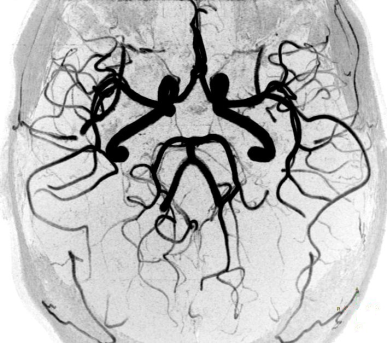
This maximum intensity projection (MIP)in the coronal plane shows the brain vessel anatomy and blood supply. The internal carotid arteries (ICA - right and left thick vessels) and the vertebral arteries join together at the base of the brain. The connection of the vertebral arteries forms the basilar artery (seen in the lower, middle part of the picture).
 
with enhanced brightness and contrast
 
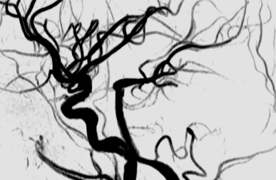
The MIP in transverse projection shows the circle of Willis, formed around the optic chiasm and the pituitary stalk (brain tissue is not seen due to MIP processing).Seen are the anterior communicating arteries (right and left ICA, right and left anterior cerebral arteries (ACA), anterior communicating artery (ACOM)) and the posterior communicating arteries (right and left posterior communicating arteries (PCOM), right and left posterior cerebral arteries (PCA), basilar artery).
 
with enhanced brightness and contrast
 
The MIP in sagittal projection shows a lateral view of the circle of Willis. The maximum intensity projections of this magnetic resonance angiography (MRA) are inverted to provide high vessel contrast. High field MRI (3T) provides the high signal to noise ratio and spatial resolution that are important when imaging flow within the intracranial vasculature.
 
with enhanced brightness and contrast
 
The used 3D time of flight angiography (TOF) provides the imaging of the arteries due to presaturation of the veins (selective MRA). TOF uses the inflow effect of gradient echo sequences to depict flowing blood as bright signal due to flow related enhancement. A short repetition time (TR) provides the saturation of the background tissue. Small voxel size (around 0,4 mm x 0,5 mm and slice thickness 0,5 mm) and a short echo time (TE) are important for minimal signal loss due to voxel dephasing (from complex or turbulent flow).
 
with enhanced brightness and contrast
 
 
Related Images
 • Brain MRI Inversion Recovery
 • Brain MRI Coronal FLAIR 001
 • Brain MRI Sagittal T1 001
 • Brain MRI Transversal T1 001
 • Brain MRI Transversal T1 002
 • Brain MRI Transversal T2 001
 • Brain MRI Transversal T2 002
 • Brain MRI Transversal T2 003
 • Brain MRI Images Axial T2
 • Brain MRI Images T1
 • PCA-MRA 3D Brain Venography Colored MIP
Related Sliders
 • MRI of the Skull Base
 • MRI of the Brain Stem with Temoral Bone and Auditory System
 • Circle of Willis, Time of Flight, MIP


Relevant Database Entries
 • Angiography
 • Brain MRI
 • Circle of Willis
 • Entry Slice Phenomenon (Artifact)
 • Flow
 • Flow Effects
 • Flow Related Enhancement
 • Inflow Magnetic Resonance Angiography
 • Magnetic Resonance Angiography MRA
 • Maximum Intensity Projection
 • MRI
 • Postprocessing
 • Spin Phase Effect
 • Time of Flight Angiography
 • Vessel Separation
 
  Next Image - PCA-MRA 3D Brain Venography Colored MIP top
You can fool some of the people all of the time, and all of the people some of the time, but you can not fool all of the people all of the time.
- Abraham Lincoln
 
Share This Page
FacebookTwitterLinkedIn

MR-TIP    
Community   
User
Pass
Forgot your UserID/Password ?    


Look
      Ups


MR-TIP.com uses cookies! By browsing MR-TIP.com, you agree to our use of cookies.

Magnetic Resonance - Technology Information Portal
Member of SoftWays' Medical Imaging Group - MR-TIP • Radiology-TIP • Medical-Ultrasound-Imaging • 
Copyright © 2003 - 2024 SoftWays. All rights reserved. [ 18 March 2024]
Terms of Use | Privacy Policy | Advertising
 [last update: 2024-02-26 03:41:00]