Magnetic Resonance - Technology Information Portal Welcome to MRI Technology
Info
  Sheets

Out-
      side
 

New acceleration techniques will :
reduce scan times 
cause artifacts 
increase expenses 
be useful if you have a lot of experience 
doesn't do much 
never heard of 


MRI Forum: Muscle shading in 3T Images
SEARCH FORUM FOR   
Forum Overview

  Category:  Applications and Examinations
Reply to this thread
(login or register first)
What is - 
'Forum Notification Service'?  
Travis Conley

Thu. 21 Oct.10,
17:22
Muscle shading in 3T Images
We are using a 3T GE scanner to image axial slices of human thigh muscle for the purpose of quantifying muscle volume. rnrnProblem: Our images have been presenting some shading at the top and bottom of each slice. We have tried everything we can think of to get rid of this shading but have not been sucessful. rnrnI have attached a representative image and would love some feel back. rnrnThanks so much for attempting to help us out.
 
 
Thigh Axial Slice


Don LaVille

Tue. 21 Dec.10,
20:57
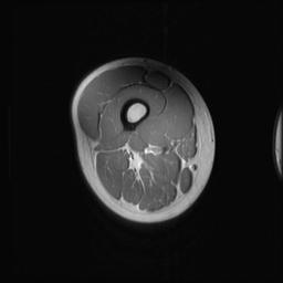
This artifact is known as the dielectric effect. It is a phenomena to all magnets. At 1.5T the dielectric effect is at 60cm so when we scan at a FOV of 40 or less we don't see it. At 3t the dielectric effect is reduced to 30cm so we now see it when using a FOV of 30 or more. There is no way to get rid of it that I know of and you will see an increase with patients with more magnetic suseptibility (ie. patients with ascities, and pregnant patient). You could try using Surface Coil Intensity Correction (SCIC) and it will help to even out the extreme dark and light in your image. Some vendors will suggest using a Fat Sat pad to help reduce this artifact. I have tried it with little success.
Looking at your image, it looks like you were scanning on a GE 3T HDX. I hope that the vendors will have a fix for this artifact soon. For right now though we are just dealing with it. I hope this helps.

Desiree Dupuy

Sat. 3 Dec.16,
22:14
Try running a B1 Map followed by Calibration. Set RF Drive mode to Optimized. This is supposed to optimize for your Patient. Supposed to reduce dielectric effect/shading for Lumbar and Abdomen.
You can find under GE Additional Abdomen Sequences.

Or you can run Ideal with Dixon technique. Turn on IN phase will give you T1 FS & IN Phase is you plane T1. I believe this takes care of the artifact.

Forum Overview

  Category:  Applications and Examinations
Reply to this thread
(login or register first)
What is - 
'Forum Notification Service'?  
Human history becomes more and more a race between education and catastrophe.
- H. G. Wells
 
Share This Page
FacebookTwitterLinkedIn

MR-TIP    
Community   
User
Pass
Forgot your UserID/Password ?    


Look
      Ups


MR-TIP.com uses cookies! By browsing MR-TIP.com, you agree to our use of cookies.

Magnetic Resonance - Technology Information Portal
Member of SoftWays' Medical Imaging Group - MR-TIP • Radiology-TIP • Medical-Ultrasound-Imaging • 
Copyright © 2003 - 2024 SoftWays. All rights reserved. [ 20 April 2024]
Terms of Use | Privacy Policy | Advertising
 [last update: 2024-02-26 03:41:00]