 | Info
Sheets |
| | | | | | | | | | | | | | | | | | | | | | | | |
 | Out-
side |
| | | | |
|
| | | | |
Result : Searchterm 'flair' found in 0 term [ ] and 49 definitions [ ] and 49 definitions [ ] ]
| | previous 41 - 45 (of 49) nextResult Pages : [1 2 3 4 5 6 7 8 9 10] |  | |  | Searchterm 'flair' was also found in the following services: | | | | |
| |  |
| |
|

From GE Healthcare;
'EXCITE technology has the potential to open the door to new imaging techniques and clinical applications, leaping beyond conventional two and three-dimensional MRI to true 4D imaging that will improve the diagnosis of disease throughout the human body from head to foot.' Robert R. Edelman, M.D., Professor of Radiology at Northwestern University Medical School and Chairman, Department of Radiology, at Evanston Northwestern Healthcare.
Device Information and Specification CLINICAL APPLICATION Whole body Head and body coil standard; all other coils optional; open architecture makes system compatible with a wide selection of coils Optional 2D/3D brain and prostate Standard: SE, IR, 2D/3D GRE and SPGR, Angiography: 2D/3D TOF, 2D/3D Phase Contrast;; 2D/3D FSE, 2D/3D FGRE and FSPGR, SSFP, FLAIR, EPI, optional: 2D/3D Fiesta, FGRET, Spiral, TensorTR 1.3 to 12000 msec in increments of 1 msec TE 0.4 to 2000 msec in increments of 1 msec 2D 0.7 mm to 20 mm; 3D 0.1 mm to 5 mm 128x512 steps 32 phase encode 0.08 mm; 0.02 mm optional POWER REQUIREMENTS 480 or 380/415 less than 0.03 L/hr liquid heliumSTRENGTH SmartSpeed 23 mT/m, HiSpeed Plus 33 mT/m, EchoSpeed Plus 33 mT/m 4.0 m x 2.8 m axial x radial | | | | | |
| | | | | |
| |
|
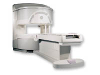
From GE Healthcare;
a friendly and less confining appearance targets the 7% of individuals who refuse to have an MRI because of claustrophobia. This open MRI system is also up to three times faster than other scanners, therefore the Signa OpenSpeed™ reducing exam time and scheduling
issues. In addition, a swing table provides better access and supports up to 500 pounds.
Device Information and Specification CLINICAL APPLICATION Whole body Standard: SE, IR, 2D/3D GRE and SPGR, Angiography: 2D/3D TOF, 2D/3D Phase Contrast;; 2D/3D FSE, 2D/3D FGRE and FSPGR, SSFP, FLAIR, EPI, optional: 2D/3D Fiesta, FGRET, Spiral, TensorTR 1.3 to 12000 msec in increments of 1 msec TE 0.4 to 2000 msec in increments of 1 msec 2D: 0.8mm - 20mm 3D: 0.1mm - 20mm 0.08 mm; 0.02 mm optional POWER REQUIREMENTS 200 - 480, 3-phase | | | |
• View the DATABASE results for 'Signa OpenSpeed™' (2).
| | | | |  Further Reading: Further Reading: | News & More:
|
|
| |
| | | | | |
| |
|
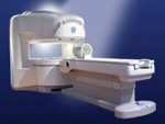
From GE Healthcare;
the Signa Ovation™ is a patient-friendly open MRI scanner designed not only to handle a typical patient mix, but to accommodate larger patients, patients who are claustrophobic, and others who have difficulty tolerating the close quarters of conventional MR machines.
Device Information and Specification CLINICAL APPLICATION Whole body Standard: SE, IR, 2D/3D GRE and SPGR, 2D/3D TOF, 2D/3D FSE, 2D/3D FGRE and FSPGR, SSFP, FLAIR, EPI, optional: 2D/3D Fiesta, true chem sat, fat/water separation, single shot diffusion EPI, line scan diffusionIMAGING MODES Localizer, single slice, multislice, volume, fast, POMP, multi slab, cine, slice and frequency zip, extended dynamic range, tailored RF TR 1.3 to 12000 msec in increments of 1 msec TE 0.4 to 2000 msec in increments of 1 msec 2D: 1.4mm - 20mm 3D: 0.2mm - 20mm 0.08 mm; 0.02 mm optional POWER REQUIREMENTS 200 - 480, 3-phase MAX. GRADIENT AMPLITUDE 19 mT/m | | | |
• View the DATABASE results for 'Signa Ovation™' (2).
| | | | |
| | | Searchterm 'flair' was also found in the following services: | | | | |
| | |
| |
|

From GE Healthcare;
The Signa SP 0.5T™ is an open MRI magnet that is designed for use in interventional radiology and intra-operative imaging. The vertical gap configuration increases patient positioning options, improves patient observation, and allows continuous access to the patient during imaging.
The magnet enclosure also incorporates an intercom, patient observation video camera, laser patient alignment lights, and task lighting in the imaging volume.
Device Information and Specification CLINICAL APPLICATION Whole body Integrated transmit and receive body coil; optional rotational body coil, head; other coils optional; open architecture makes system compatible with a wide selection of coilsarray Standard: SE, IR, 2D/3D GRE and SPGR, 2D/3D TOF, 2D/3D FSE, 2D/3D FGRE and FSPGR, SSFP, FLAIR, EPI, optional: 2D/3D Fiesta, true chem sat, fat/water separation, single shot diffusion EPI IMAGING MODES Localizer, single slice, multislice, volume, fast, POMP, multi slab, cine, slice and frequency zip, extended dynamic range, tailored RF TR 1.3 to 12000 msec in increments of 1 msec TE 0.4 to 2000 msec in increments of 1 msec 2D: 1.4mm - 20mm 3D: 0.2mm - 20mm POWER REQUIREMENTS 200 - 480, 3-phase | | | |
• View the DATABASE results for 'Signa SP 0.5T™ Open Configuration' (2).
| | | | | | Further Reading: | News & More:
|
|
| |
| | | | | |
| |
|
Magnetic resonance imaging ( MRI) of the spine is a noninvasive procedure to evaluate different types of tissue, including the spinal cord, vertebral disks and spaces between the vertebrae through which the nerves travel, as well as distinguish healthy tissue from diseased tissue.
The cervical, thoracic and lumbar spine MRI should be scanned in individual sections.
The scan protocol parameter like e.g. the field of view ( FOV), slice thickness and matrix are usually different for cervical, thoracic and lumbar spine MRI, but the method
is similar. The standard views in the basic spinal MRI scan to create detailed slices (cross sections) are sagittal T1 weighted and T2 weighted images over the whole body part, and transverse (e.g. multi angle oblique) over the region of interest with different pulse sequences according to the result of the sagittal slices. Additional views or different types of pulse sequences like fat suppression, fluid attenuation inversion recovery ( FLAIR) or
diffusion weighted imaging are created dependent on the indication.
Indications:
•
Neurological deficit, evidence of radiculopathy, cauda equina compression
•
Primary tumors or drop metastases
•
Infection/inflammatory disease, multiple sclerosis
•
Postoperative evaluation of lumbar spine: disk vs. scar
•
Localized back pain with no radiculopathy (leg pain)
Contrast enhanced MRI techniques delineate infections vs. malignancies, show a syrinx cavity and support to differentiate the postoperative conditions. After surgery for disk disease, significant fibrosis can occur in the spine. This scarring can mimic residual disk herniation. Magnetic resonance myelography evaluates spinal stenosis and various intervertebral discs can be imaged with multi angle oblique techniques. Cine series can be used to show true range of motion studies of parts of the spine.
Advanced open MRI devices are developed to perform positional scans in the position of pain or symptom (e.g. Upright™ MRI formerly Stand-Up MRI). | | | | | |
• View the DATABASE results for 'Spine MRI' (11).
| | |
• View the NEWS results for 'Spine MRI' (4).
| | | | | | Further Reading: | | Basics:
|
|
News & More:
| |
| |
| | | | |
| | |
| | | |
|
| |
 | Look
Ups |
| |


