 | Info
Sheets |
| | | | | | | | | | | | | | | | | | | | | | | | |
 | Out-
side |
| | | | |
|
| | | | | |  |
Result : Searchterm 'flair' found in 0 term [ ] and 49 definitions [ ] and 49 definitions [ ] ]
| | 1 - 5 (of 49) nextResult Pages : [1 2 3 4 5 6 7 8 9 10] | | |  | Searchterm 'flair' was also found in the following services: | | | | |
| |  |
| |
|
| | | | | | | | • Share the entry 'Fluid Attenuation Inversion Recovery':    | | | | | | | | | |  Further Reading: Further Reading: | | Basics:
|
|
News & More:
| |
| |
| | | | | |
| |
|
Brain imaging, magnetic resonance imaging of the head or skull, cranial magnetic resonance tomography (MRT), neurological MRI - they describe all the same radiological imaging technique for medical diagnostic.
Magnetic resonance imaging of the human brain includes the anatomic description and the detection of lesions. Special techniques like diffusion weighted imaging, functional magnetic resonance imaging ( fMRI) and spectroscopy provide also information about the function and chemical metabolites of the brain.
MRI provides detailed pictures of brain and nerve tissues in multiple planes without obstruction by overlying bones. Brain MRI is the procedure of choice for most brain disorders. It provides clear images of the brainstem and posterior brain, which are difficult to view on a CT scan. It is also useful for the diagnosis of demyelinating disorders (disorders such as multiple sclerosis (MS) that cause destruction of the myelin sheath of the nerve).
With this noninvasive procedure also the evaluation of blood flow and the flow of cerebrospinal fluid (CSF) is possible. Different MRA methods, also without contrast agents can show a venous or arterial angiogram. MRI can distinguish tumors, inflammatory lesions, and other pathologies from the normal brain anatomy. However, MRI scans are also used instead other methods to avoid the dangers of interventional procedures like angiography (DSA - digital subtraction angiography) as well as of repeated exposure to radiation as required for computed tomography (CT) and other X-ray examinations.
A ( birdcage) bird cage coil achieves uniform excitation and reception and is commonly used to study the brain. Usually a brain MRI procedure includes FLAIR, T2 weighted and T1 weighted sequences in two or three planes. See also Fetal MRI, Fluid Attenuation Inversion Recovery ( FLAIR), Perfusion Imaging and High Field MRI. See also Arterial Spin Labeling. | | | | | | | |
• View the DATABASE results for 'Brain MRI' (14).
| | |
• View the NEWS results for 'Brain MRI' (32).
| | | | | | Further Reading: | Basics:
|
|
News & More:
|  |
MRI Reveals Significant Brain Abnormalities Post-COVID
Monday, 21 November 2022 by neurosciencenews.com | | |
Combining genetics and brain MRI can aid in predicting chances of Alzheimer's disease
Wednesday, 29 June 2022 by www.sciencedaily.com | | |
Roundup: How Even Mild COVID Can Affect the Brain; This Many Daily Steps Improves Longevity; and More
Friday, 11 March 2022 by baptisthealth.net | | |
A low-cost and shielding-free ultra-low-field brain MRI scanner
Tuesday, 14 December 2021 by www.nature.com | | |
Large International Study Reveals Spectrum of COVID-19 Brain Complications
Tuesday, 9 November 2021 by www.itnonline.com | | |
Brain MRI-Based Subtypes of MS Predict Disability Progression, Treatment Response
Thursday, 13 May 2021 by www.neurologyadvisor.com | | |
New MRI method improves detection of disease changes in the brain's network
Thursday, 11 June 2020 by www.compute.dtu.dk | | |
New NeuroCOVID Classification System Uses MRI to Categorize Patients
Friday, 12 June 2020 by www.diagnosticimaging.com | | |
New MRI technique can 'see' molecular changes in the brain
Thursday, 5 September 2019 by medicalxpress.com | | |
Talking therapy or medication for depression: Brain scan may help suggest better treatment
Monday, 27 March 2017 by www.newsnation.in | | |
MRI identifies brain abnormalities in chronic fatigue syndrome patients
Wednesday, 29 October 2014 by www.eurekalert.org | | |
MRIs Useful in Tracking Depression in MS Patients
Tuesday, 1 July 2014 by www.hcplive.com | | |
Contrast agent linked with brain abnormalities on MRI
Tuesday, 17 December 2013 by www.sciencecodex.com | | |
MRIs Reveal Signs of Brain Injuries Not Seen in CT Scans
Tuesday, 18 December 2012 by www.sciencedaily.com | | |
Iron Deposits in the Brain May Be Early Indicator of MS
Wednesday, 13 November 2013 by www.healthline.com | | |
Migraine Sufferers Have Thicker Brain Cortex
Tuesday, 20 November 2007 by www.medicalnewstoday.com |
|
| |
| | | | | |
| |
|

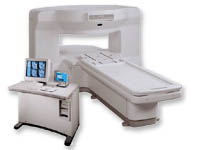
'MRI system is not an expensive equipment anymore.
ENCORE developed by ISOL Technology is a low cost MRI system with the advantages like of the 1.0T MRI scanner. Developed specially for the overseas market, the ENCORE is gaining popularity in the domestic market by medium sized hospitals.
Due to the optimum RF and Gradient application technology. ENCORE enables to obtain high resolution imaging and 2D/3D Angio images which was only possible in high field MR systems.'
- Less consumption of the helium gas due to the ultra-lightweight magnet specially designed and manufactured for ISOL.
- Cost efficiency MR system due to air cooling type (equivalent to permanent magnetic).
- Patient processing speed of less than 20 minutes.'
Device Information and Specification
CLINICAL APPLICATION
Whole body
CONFIGURATION
Short bore compact
| | | |
• View the DATABASE results for 'ENCORE 0.5T™' (2).
| | | | |
| | | Searchterm 'flair' was also found in the following services: | | | | |
| | |
| |
|

From Philips Medical Systems;
this active shielded member of the Panorama product line combines the advantages of one 1.0 T system's with the possibilities of an open MRI system. The open design helps ease anxiety for claustrophobic patients and increased patient comfort whereby the field strength provides spectacular image quality and fast patient throughput.
Device Information and Specification CLINICAL APPLICATION Whole body Vertically opposed solenoids, head, head-neck, extremity, neck, body/ spine M-XL, shoulder, bilateral breast, wrist, TMJ, flex XS-S-M-L-XL-XXL SE, FE, IR, STIR, FFE, DEFFE, DESE, TSE, DETSE, Single shot SE, DRIVE, Balanced FFE, MRCP, FLAIR, Turbo FLAIR, IR-TSE, T1-STIR TSE, T2-STIR TSE, Diffusion Imaging, 3D SE, 3D FFE, Contrast Perfusion Analysis, MTC;; Angiography: CE-ANGIO, MRA 2D, 3D TOFOpen x 47 cm x infinite (side-first patient entry) POWER REQUIREMENTS 400/480 V | | | |
• View the DATABASE results for 'Panorama 1.0T™' (2).
| | | | |
| | | | | |
| |
|
From GE Healthcare;
the New Signa Profile/i is a patient friendly open MRI system that virtually eliminates patient anxiety and claustrophobia, without compromising diagnostic utility.
Device Information and Specification CLINICAL APPLICATION Whole body Integrated transmit body coil, body flex sizes: M, L, XL, quadrature, head coil quadrature, 4 channel neurovascular array, 8 channel CTL array, quad. c- spine, 2 channel shoulder array, extremity coil, 3 channel wrist array, 4 channel breast array, 6, 9, 11 inch general purpose loop coils Standard: SE, IR, 2D/3D GRE and SPGR, Angiography: 2D/3D TOF, 2D/3D phase contrast; 2D/3D FSE, 2D/3D FRFSE, FGRE and FSPGR, SSFP, FLAIR, EPI, optional: 2D/3D Fiesta, fat/water separation, T1 FLAIRIMAGING MODES Localizer, single slice, multislice, volume, fast, POMP, multi slab, cine, slice and frequency zip, extended dynamic range, tailored RF TR 6 to 12000 msec in increments of 1 msec TE 1.3 to 2000 msec in increments of 1 msec 2D: 2.7mm - 20mm 3D: 0.2mm - 5mm 0.08 mm; 0.02 mm optional 10,000 kg w/gradient enclosure POWER REQUIREMENTS 200 - 480, 3-phase COOLING SYSTEM TYPE None required | | | |
• View the DATABASE results for 'Signa Profile™' (2).
| | | | |
| | | | |
| | | |
|
| |
 | Look
Ups |
| |


