 | Info
Sheets |
| | | | | | | | | | | | | | | | | | | | | | | | |
 | Out-
side |
| | | | |
|
| | | | |
Result : Searchterm 'Vantage' found in 1 term [ ] and 68 definitions [ ] and 68 definitions [ ] ]
| | 1 - 5 (of 69) nextResult Pages : [1] [2 3 4 5 6 7 8 9 10 11 12 13 14] |  | |  | Searchterm 'Vantage™' was also found in the following service: | | | | |
| |  |
| |
|
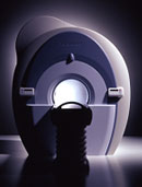
From Toshiba America Medical Systems Inc.;
With its high-field strength, the Vantage™ delivers the clinical capabilities and image quality expected by cardiologists, while simultaneously offering patients a more comfortable and non-invasive option, said Dane Peshe, director, MRI Business Unit, Toshiba America Medical Systems. Vantage™ supports a full complement of cardiovascular imaging studies, ranging from stroke evaluation to peripheral vascular imaging. Additionally, the ultra short bore design offers patients a greater feeling of openness that reduces claustrophobic sensations, while Toshiba's exclusive, patented Pianissimo™ technology reduces scan noise by as much as 90 percent for a more pleasant experience.'
Device Information and Specification CLINICAL APPLICATION Whole body CONFIGURATION Ultra short bore SE, FE, IR, FastSE, FastIR, FastFLAIR, Fast STIR, FastFE, FASE, EPI, SuperFASE; Angiography: 2D(gate/non-gate)/3D TOF, SORS-STC IMAGING MODES Single, multislice, volume study 32-1024, phase;; 64-1024, freq. POWER REQUIREMENTS 380/400/415/440/480 V COOLING SYSTEM TYPE Closed-loop water-cooled Liquid helium: approx. less than 0.05 L/hr Passive, active, auto-active | | | | | | • Share the entry 'Vantage™':    | | | | |  Further Reading: Further Reading: | Basics:
|
|
| |
| | | | |  |
| |
|
A RF coil, often a transmit receive coil with a number of wires running along the z-direction, arranged to give a cosine current variation around the circumference of the coil, which looks like a bird cage.
The bird cage coil works on a different principle to conventionally tuned local and surround coils in that it behaves like a tuned transmission line with one complete cycle of standing wave around the circumference. The frequency supply is generated by an oscillator, which is modulated to form a shaped pulse by a product detector controlled by the waveform generator. The signal must be amplified to 1000's of watts. This can be done using either solid state electronics, valves or a combination of both.
The bird cage coil design provides the best field homogeneity of all RF imaging coils.
One ad vantage is that it is simple to produce an exceedingly uniform B1 radio frequency field over most of the coil's volume, with the result of images with a high degree of uniformity.
A second ad vantage is that nodes with zero voltage occur 90° away from the driven part of the coil, thus facilitating the introduction of a second signal in quadrature, which produces a circularly polarized radio frequency field.
This type of volume coil is used for brain (head) MRI, or MR imaging of joints, such as the wrist or knees.
See also the related poll result: ' 3rd party coils are better than the original manufacturer coils' | | | | | |
• View the DATABASE results for 'Bird Cage Coil' (4).
| | | | | | Further Reading: | | Basics:
|
|
News & More:
| |
| |
| | | | | |
| |
|
| | | |
• View the DATABASE results for 'Blood Pool Agents' (16).
| | |
• View the NEWS results for 'Blood Pool Agents' (1).
| | | | | | Further Reading: | | Basics:
|
|
News & More:
| |
| |
| | | Searchterm 'Vantage™' was also found in the following service: | | | | |
| | |
| |
|
Burst pulse sequences are fast imaging sequences capable of image acquisition in less than 100 ms.
Basically a train of low flip angle pulses generates a long train of echoes. The complete sequence is performed with the application of a constant read gradient. Phase encoding may be implemented using short phase encoding gradients between echoes.
The ad vantage of this sequence type is that it is less demanding on gradient speed than other fast techniques (e.g. echo planar imaging EPI) and it produces images, which are substantially free of susceptibility artifacts.
The disad vantage is that the technique is less sensitive than competing methods. | | | | | |
| | | | | |
| |
|
(CE MRA) Contrast enhanced MR angiography is based on the T1 values of blood, the surrounding tissue, and paramagnetic contrast agent.
T1-shortening contrast agents reduces the T1 value of the blood (approximately to 50 msec, shorter than that of the surrounding tissues) and allow the visualization of blood vessels, as the images are no longer dependent primarily on the inflow effect of the blood.
Contrast enhanced MRA is performed with a short TR to have low signal (due to the longer T1) from the stationary tissue, short scan time to facilitate breath hold imaging, short TE to minimize T2* effects and a bolus injection of a sufficient dose of a gadolinium chelate.
Images of the region of interest are performed with 3D spoiled gradient echo pulse sequences. The enhancement is maximized by timing the contrast agent injection such that the period of maximum arterial concentration corresponds to the k-space acquisition. Different techniques are used to ensure optimal contrast of the arteries e.g., bolus timing, automatic bolus detection, bolus tracking, care bolus.
A high resolution with near isotropic voxels and minimal pulsatility and misregistration artifacts should be striven for. The postprocessing with the maximum intensity projection ( MIP) enables different views of the 3D data set.
Unlike conventional MRA techniques based on velocity dependent inflow or phase shift techniques, contrast enhanced MRA exploits the
gadolinium induced T1-shortening effects. CE MRA reduces or eliminates most of the artifacts of time of flight angiography or phase contrast angiography. Ad vantages are the possibility of in plane imaging of the blood vessels, which allows to examine large parts in a short time and high resolution scans in one breath hold.
CE MRA has found a wide acceptance in the clinical routine, caused by the
ad vantages:
•
3D MRA can be acquired in any plane, which means that
greater vessel coverage can be obtained at high
resolution with fewer slices (aorta, peripheral vessels);
•
the possibility to perform a time resolved examination
(similarly to conventional angiography);
•
no use of ionizing radiation; paramagnetic agents have a beneficial safety.
| | | | | |
• View the DATABASE results for 'Contrast Enhanced Magnetic Resonance Angiography' (14).
| | |
• View the NEWS results for 'Contrast Enhanced Magnetic Resonance Angiography' (2).
| | | | | | Further Reading: | Basics:
|
|
News & More:
| |
| |
| | | | |
| | | |
|
| |
 | Look
Ups |
| |


