 | Info
Sheets |
| | | | | | | | | | | | | | | | | | | | | | | | |
 | Out-
side |
| | | | |
|
| | | | | |  | Searchterm 'Spin' was also found in the following services: | | | | |
|  |  |
| |
|
The T2 time constant, which determines the rate at which excited protons reach equilibrium, or go out of phase with each other. A measure of the time taken for spinning protons to lose phase coherence among the nuclei spinning perpendicular to the main field due to interaction between spins, resulting in a reduction in the transverse magnetization. The transverse magnetization value will drop from maximum to a value of about 37% of its original value in a time of T2. | | | |  | | | |
|  |  | Searchterm 'Spin' was also found in the following services: | | | | |
|  |  |
| |
|
| |  | | | |  Further Reading: Further Reading: | News & More:
|
|
| |
|  | |  |  |  |
| |
|
Inert hyperpolarized gases are under development for imaging air spaces, including those in the lungs. Because they mostly contain air and water, lungs are difficult organs to image.
These ventilation agents (gases) have potential in lung imaging and are currently used in studies of the pulmonary ventilation:
•
aerosolized gadolinium-DTPA
•
hyperpolarized gases (xenon-129, helium-3)
Specific isotopes of inert gases can be hyperpolarized. Hyperpolarized is a state in which almost all of the atoms nuclei are spinning in the same direction. Once the nuclei in the isotope 3He have been hyperpolarized using a laser, they remain in this state for several days.
The inert, hyperpolarized gas can then be used in a lung imaging study, where the high concentration of polarized nuclei provides a sharp contrast in MRI. The technique is already being developed with a view to commercialization by Magnetic Imaging Technologies in Durham, North Carolina. According to the company, existing MRI equipment can be used with a few minor modifications, along with a gas polarizer. The technique could provide early detection and monitoring of pulmonary disease.
Hyperpolarized 129Xe can also be used as a magnetic resonance tracer because of its MR-enhanced sensitivity combined with its high solubility.
This isotope differs from 3He in that it can dissolve in the blood. Strong enhancement of the nuclear spin polarization of xenon in the gas phase can be achieved by optical pumping of rubidium and subsequent spin-exchange with the xenon nuclei.
This technique can increase the magnetic resonance signal of xenon by five orders of magnitude, thus allowing NMR detection of xenon in very low concentration. MR spectroscopy and imaging of optically polarized xenon shows considerable potential for medical applications (see also back projection imaging).
Nycomed Amersham anticipated the market for inert gases in pulmonary imaging. The company obtained an exclusive license for the use of helium (He) and xenon (Xe) as MRI contrast agents. Currently, the US FDA has not yet approved the commercial distribution of inert gas imaging equipment, because the technique is still undergoing trials. | |  | |
• View the DATABASE results for 'Ventilation Agents' (3).
| | | | |  Further Reading: Further Reading: | | Basics:
|
|
News & More:
| |
| |
|  |  | Searchterm 'Spin' was also found in the following services: | | | | |
|  |  |
| |
|

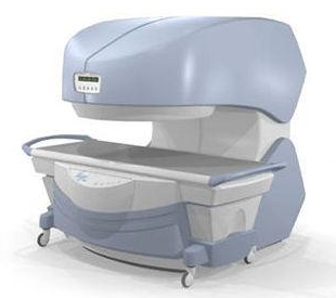
From
Millennium Technology Inc.
This open C-shaped MRI system eases patient comfort and technologist maneuverability. This low cost scanner is build for a wide range of applications. The Virgo™ patient table is detachable and moves on easy rolling castors. Able to accommodate patient weights up to 160 kg, the tabletop has a range of motion of 30 cm in the lateral direction and 90cm in the longitudinal direction. Images generated with this scanner can only be viewed (without data loss) on Millennium's proprietary viewing software.
Device Information and Specification CLINICAL APPLICATION Whole body Head, Body, Neck, Knee, Shoulder,
Spine, Wrist, Breast, Extremity, Lumbar Spine, TMJ
IMAGING MODES Localizer, single slice, multislice, volume, fast, POMP, multi slab, cine, slice and frequency zip, extended dynamic range, tailored RF | |  | | | |
|  |  | Searchterm 'Spin' was also found in the following services: | | | | |
|  |  |
| |
|

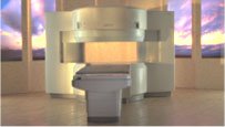
From Hitachi Medical Systems America Inc.;
the AIRIS II, an entry in the diagnostic category of open MR systems, was designed by Hitachi
Medical Systems America Inc. (Twinsburg, OH, USA) and Hitachi Medical Corp. (Tokyo) and is manufactured by the Tokyo branch. A 0.3 T field-strength magnet and phased array coils deliver high image quality without the need for a tunnel-type high-field system, thereby significantly improving patient comfort not only for claustrophobic patients.
Device Information and Specification
CLINICAL APPLICATION
Whole body
QD Head, MA Head and Neck, QD C-Spine, MA or QD Shoulder, MA CTL Spine, QD Knee, Neck, QD TMJ, QD Breast, QD Flex Body (4 sizes), Small and Large Extrem., QD Wrist, MA Foot and Ankle (WIP), PVA (WIP)
SE, GE, GR, IR, FIR, STIR, FSE, ss-FSE, FLAIR, EPI -DWI, SE-EPI, ms - EPI, SSP, MTC, SARGE, RSSG, TRSG, MRCP, Angiography: CE, 2D/3D TOF
IMAGING MODES
Single, multislice, volume study
TR
SE: 30 - 10,000msec GE: 20 - 10,000msec IR: 50 - 16,700msec FSE: 200 - 16,7000msec
TE
SE : 10 - 250msec IR: 10 -250msec GE: 5 - 50 msec FSE: 15 - 2,000
0.05 sec/image (256 x 256)
2D: 2 - 100 mm; 3D: 0.5 - 5 mm
Level Range: -2,000 to +4,000
POWER REQUIREMENTS
208/220/240 V, single phase
COOLING SYSTEM TYPE
Air-cooled
2.0 m lateral, 2.5 m vert./long
| |  | |
• View the DATABASE results for 'AIRIS II™' (2).
| | | | |
|  | |  |  |
|  | | |
|
| |
 | Look
Ups |
| |