Magnetic Resonance - Technology Information Portal Welcome to MRI Technology
Info
  Sheets

Out-
      side


 'Slice' 
SEARCH FOR    
 
  2 3 5 A B C D E F G H I J K L M N O P Q R S T U V W X Y Z
Result : Searchterm 'Slice' found in 19 terms [] and 159 definitions []
previous     31 - 35 (of 178)     next
Result Pages : [1 2 3 4]  [5 6 7 8 9 10 11 12 13 14 15 16 17 18 19 20 ... ]
Searchterm 'Slice' was also found in the following services: 
spacer
News  (6)  Resources  (5)  Forum  (25)  
 
Staircase ArtifactInfoSheet: - Artifacts - 
Case Studies, 
Reduction Index, 
etc.MRI Resource Directory:
 - Artifacts -
 
Quick Overview
Artifact Information
NAME
Staircase
DESCRIPTION
Staircase like nuance
REASON
Slices too thick, no overlapping
HELP
Overlapping slices
Staircase artifacts might be seen in projection of the slice thickness in case of MIP or MPR reconstructions.
mri safety guidance
Image Guidance
Use overlapping slices to reduce this artifact by changing the slice gap to a negative value. In most cases, a negative value of 20-30% is sufficient.
spacer
Searchterm 'Slice' was also found in the following services: 
spacer
Radiology  (24) Open this link in a new windowUltrasound  (9) Open this link in a new window
Upright™ MRIInfoSheet: - Devices -
Intro, 
Types of Magnets, 
Overview, 
etc.MRI Resource Directory:
 - Devices -
 
www.fonar.com/standup.htm www.fonar.com/standup.htm www.fonar.com/standup.htm 
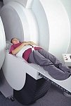
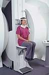
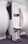
From FONAR Corporation;
in October of 2004, the company changed the product name of the Stand-Up™ MRI to the Upright™ MRI. The Indomitable™, Upright™ MRI is the only open MRI in the world that can perform positional MRI (pMRI), i.e. the Upright MRI™ scans patients in upright, weight-bearing positions, in addition to the conventional lie-down positions. The Upright™ MRI is the only device that can scan patients in the position their symptoms occur, in their position of pain. In early clinical reports independently confirm the effectiveness and potential of positional MRI. In October 2000, Fonar received permission to market the Indomitable™ from the FDA.
Device Information and Specification
CLINICAL APPLICATION
Whole body - weight-bearing MRI - position imaging (flexion, extension, bending, standing, sitting and recumbent scanning)
CONFIGURATION
Front-open and Top-open MRI
Solenoid ("Wrap-around") and planar RF receiver coils
SINGLE/MULTI SLICE
0.1 sec per slice
MULTISLICE
0.1 sec per slice
Min. 6.0 cm
Min. 0.8 mm (3DFT)
MEASURING MATRIX
128 - 512
MAGNET TYPE
Iron-frame electromagnet
45.72 cm horizontal
POWER REQUIREMENTS
380/400/415/440/480 V
COOLING SYSTEM TYPE
Water, closed-loop
CRYOGEN USE, L/hr
-
STRENGTH
12 mT/m
5 GAUSS FRINGE FIELD, radial/axial
active, passive
spacer

• View the DATABASE results for 'Upright™ MRI' (5).Open this link in a new window


• View the NEWS results for 'Upright™ MRI' (9).Open this link in a new window.
 
Further Reading:
  Basics:
Product Information - Fonar Corporation
  News & More:
Landmark Independent Study by UCLA School of Medicine Reports Comparison of Dynamic™ Upright® MRI With Static Upright MRI in More Than 1,000 Patients (1,302):
Thursday, 15 November 2007   by www.fonar.com    
FONAR Wins The Wall Street Journal Technology Innovation Award For 2007 For the FONAR UPRIGHT™ Multi-Position™ MRI
Monday, 1 October 2007   by www.fonar.com    
MRI Resources 
Quality Advice - Implant and Prosthesis - MRI Centers - Shielding - Brain MRI - Journals
 
Virgo™InfoSheet: - Devices -
Intro, 
Types of Magnets, 
Overview, 
etc.MRI Resource Directory:
 - Devices -
 
www.canamglobal.com/mri/virgomri.html From Millennium Technology Inc. This open C-shaped MRI system eases patient comfort and technologist maneuverability. This low cost scanner is build for a wide range of applications. The Virgo™ patient table is detachable and moves on easy rolling castors. Able to accommodate patient weights up to 160 kg, the tabletop has a range of motion of 30 cm in the lateral direction and 90cm in the longitudinal direction. Images generated with this scanner can only be viewed (without data loss) on Millennium's proprietary viewing software.
Device Information and Specification
CLINICAL APPLICATION
Whole body
CONFIGURATION
C shaped
SYNCHRONIZATION
Standard cardiac gating, ECG/peripheral, respiratory gating
PULSE SEQUENCES
2D Spin Echo (single and multi-echo), 2D Inversion Recovery, 2D Sequential and 3D Volume Gradient Echo, 2D and 3D Spoiled Gradient Echo
IMAGING MODES
Localizer, single slice, multislice, volume, fast, POMP, multi slab, cine, slice and frequency zip, extended dynamic range, tailored RF
TR
steps of 1 msec
TE
steps of 1 msec
SINGLE/MULTI SLICE
Simultaneous scan and reconstruction;; 100 images/second reconstruction
400 mm
2D : 2 mm; 3D : 0.5 mm
MEASURING MATRIX
512x512
PIXEL INTENSITY
256 gray levels
MAGNET TYPE
Permanent
BORE DIAMETER
or Vertical Gap
44 cm
STRENGTH
15 mT/m
5-GAUSS FRINGE FIELD
3 m/3 m
Passive
spacer
Searchterm 'Slice' was also found in the following services: 
spacer
News  (6)  Resources  (5)  Forum  (25)  
 
ARTOSCAN™ - MInfoSheet: - Devices -
Intro, 
Types of Magnets, 
Overview, 
etc.MRI Resource Directory:
 - Devices -
 
www.gemedicalsystems.com/rad/mri/products/artoscan/specs.html Developed by GE Lunar; the ARTOSCAN™-M is designed specifically for in-office musculoskeletal imaging. ARTOSCAN-M's compact, modular design allows placing within a clinical environment, bringing MRI to the patient. Patients remain outside the magnet at all times during the examinations, enabling constant patient-technologist contact. ARTOSCAN-M requires no special RF room, magnetic shielding, special power supply or air conditioning.
The C-SCAN™ (also known as Artoscan C) is developed from the ARTOSCAN™ - M, with a new computer platform.
Device Information and Specification
CLINICAL APPLICATION
Dedicated extremity
CONFIGURATION
Closed
Linear and dual phased array coil, knee, ankle, wrist coils
PULSE SEQUENCES
SE, GE, IR, STIR, FSE, 3D CE, GE-STIR, 3D GE, ME, TME, HSE
IMAGING MODES
Single, multislice, volume study, fast scan, multi slab, cine
TR
12 - 5,000 msec
TE
5 - 220 msec
SINGLE SLICE
0.8 sec/image
MULTISLICE
0.8 sec/image
FOV
10 - 20 cm
SLICE THICKNESS
2D: 2 mm - 10 mm;
3D: 0.6 mm - 10 mm
512 x 512
MEASURING MATRIX
256 x 256 maximum
PIXEL INTENSITY
4,096 gray lvls, 256 lvls in 3D
MAGNET TYPE
Ferrite Permanent
MAGNET WEIGHT
965 kg
POWER REQUIREMENTS
100/110/200/220/230/240V
STRENGTH
10 mT/m
5 GAUSS FRINGE FIELD, radial/axial
28 cm/60 cm
Passive
spacer

• View the DATABASE results for 'ARTOSCAN™ - M' (3).Open this link in a new window

Searchterm 'Slice' was also found in the following services: 
spacer
Radiology  (24) Open this link in a new windowUltrasound  (9) Open this link in a new window
C-SCAN™InfoSheet: - Devices -
Intro, 
Types of Magnets, 
Overview, 
etc.MRI Resource Directory:
 - Devices -
 
www.gehealthcare.com/usen/mr/cscan/index.html Manufactured by Esaote S.p.A.; compact in-office MRI system, fits in a 90 ft2 (8.4 m2) space and requires no shielding or special power. This low field MRI magnet is optimized for orthopedic use and imaging of the extremities.
The C-SCAN™ is developed from the ARTOSCAN™ - M with a new computer platform, and is also known as Artoscan C.
Esaote North America and Hologic Inc. are the U.S. distributors of this MRI device.
Device Information and Specification
CLINICAL APPLICATION
Dedicated extremity
CONFIGURATION
Closed
Linear and dual phased array, knee, ankle, wrist (6 total) coils
PULSE SEQUENCES
SE, GE, IR, STIR, FSE, 3D CE, GE-STIR, 3D GE, ME, TME, HSE
IMAGING MODES
Single, multislice, volume study, fast scan, multi slab
TR
12 - 5,000 msec
TE
5 - 220 msec
SINGLE SLICE
0.8 sec/image
MULTISLICE
0.8 sec/image
FOV
10 - 20 cm
2D: 2 mm - 10 mm;
3D: 0.6 mm - 10 mm
512 x 512
MEASURING MATRIX
256 x 256 maximum
PIXEL INTENSITY
4,096 gray lvls, 256 lvls in 3D
MAGNET TYPE
Permanent
BORE DIAMETER
or W x H
33.6 x 16 cm
MAGNET WEIGHT
960 kg
H*W*D
124 x 76 x 60 cm
POWER REQUIREMENTS
100/110/200/220/230/240
STRENGTH
10 mT/m
5-GAUSS FRINGE FIELD, radial/axial
28 cm/60 cm
Passive
spacer

• View the DATABASE results for 'C-SCAN™' (4).Open this link in a new window

MRI Resources 
Portals - Absorption and Emission - Pathology - Raman Spectroscopy - DICOM - Libraries
 
previous      31 - 35 (of 178)     next
Result Pages : [1 2 3 4]  [5 6 7 8 9 10 11 12 13 14 15 16 17 18 19 20 ... ]
 Random Page
 
Share This Page
FacebookTwitterLinkedIn

MR-TIP    
Community   
User
Pass
Forgot your UserID/Password ?    


How AI will impact MRI :
only diagnostics 
saving time 
reducing cost 
makes planning obsolete 
reduce human knowledge 
not at all 

Look
      Ups


MR-TIP.com uses cookies! By browsing MR-TIP.com, you agree to our use of cookies.

Magnetic Resonance - Technology Information Portal
Member of SoftWays' Medical Imaging Group - MR-TIP • Radiology-TIP • Medical-Ultrasound-Imaging • 
Copyright © 2003 - 2024 SoftWays. All rights reserved. [ 17 April 2024]
Terms of Use | Privacy Policy | Advertising
 [last update: 2024-02-26 03:41:00]