Magnetic Resonance - Technology Information Portal Welcome to MRI Technology
Info
  Sheets

Out-
      side


 'Slice' 
SEARCH FOR    
 
  2 3 5 A B C D E F G H I J K L M N O P Q R S T U V W X Y Z
Result : Searchterm 'Slice' found in 19 terms [] and 159 definitions []
previous     96 - 100 (of 178)     next
Result Pages : [1 2 3 4]  [5 6 7 8 9 10 11 12 13 14 15 16 17 18 19 20 ... ]
Searchterm 'Slice' was also found in the following services: 
spacer
News  (6)  Resources  (5)  Forum  (25)  
 
Signa HDx 3.0T™InfoSheet: - Devices -
Intro, 
Types of Magnets, 
Overview, 
etc.
 
gehealthcare.com/euen/mri/products/signa-hdx-3t/index.html From GE Healthcare;
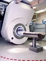
The Signa HDx MRI system is GE's leading edge whole body magnetic resonance scanner designed to support high resolution, high signal to noise ratio, and short scan times.
Signa HDx 3.0T offers new technologies like ultra-fast image reconstruction through the new XVRE recon engine, advancements in parallel imaging algorithms and the broadest range of premium applications. The HD applications, PROPELLER (high-quality brain imaging extremely resistant to motion artifacts), TRICKS (contrast-enhanced angiographic vascular lower leg imaging), VIBRANT (for breast MRI), LAVA (high resolution liver imaging with shorter breath holds and better organ coverage) and MR Echo (high-definition cardiac images in real time) offer unique capabilities.
Device Information and Specification
CLINICAL APPLICATION
Whole body
CONFIGURATION
Compact short bore
Head and body coil, T/R quadrature head; optional coils e.g., T/R phased array extremity abdomen, spine, breast, knee, shoulder, cardiac imaging coils
SYNCHRONIZATION
ECG/peripheral, respiratory gating
PULSE SEQUENCES
SE, IR, 2D/3D GRE, RF-spoiled GRE, 2DFGRE, 2DFSPGR, 3DFGRE, 3DFSPGR, 3DTOFGRE, 3DFSPGR, 2DFSE, 2DFSE-XL, 2DFSE-IR, T1-FLAIR, SSFSE, EPI, DW-EPI, BRAVO, Angiography: 2D/3D TOF, 2D/3D phase contrast vascular
IMAGING MODES
Single, multislice, volume study, fast scan, multi slab, cine, localizer
1 cm to 40 cm continuous
2D 0.5 mm; 3D 0.1 mm
1024 x 1024
PIXEL INTENSITY
256 gray levels
60 cm
MAGNET WEIGHT
12000 kg
H*W*D
240 x 2216,6 x 201,6 cm
POWER REQUIREMENTS
480 or 380/415, 3 phase ||
COOLING SYSTEM TYPE
Closed-loop water-cooled grad.
0.03 L/hr helium
STRENGTH
23 - 50 mT/m
80 - 150 mT/m/ms
5-GAUSS FRINGE FIELD
2.8 m / 5.0 m
second and high order
spacer
Searchterm 'Slice' was also found in the following services: 
spacer
Radiology  (24) Open this link in a new windowUltrasound  (9) Open this link in a new window
Spin Phase EffectInfoSheet: - Artifacts - 
Case Studies, 
Reduction Index, 
etc.
 
The spins flow with the blood through a slice and experience a RF pulse. If they flow out of the slice by the time the signal is recorded (because the repetition time (TR) is asynchronous with the pulsatile flow), the flowing blood produces intravascular signal void by 'time of flight' effects, turbulent dephasing and first echo dephasing. The liquid flow occasionally produces an intravascular high signal intensity due to flow related enhancement, even echo rephasing and diastolic pseudogating.

See also Flow Artifact and Flow Effects.
 
Images, Movies, Sliders:
 TOF-MRA Circle of Willis Inverted MIP  Open this link in a new window


spacer

• View the DATABASE results for 'Spin Phase Effect' (3).Open this link in a new window

MRI Resources 
Devices - MRA - Most Wanted - Homepages - Spine MRI - Examinations
 
StacksInfoSheet: - Sequences - 
Intro, 
Overview, 
Types of, 
etc.
 
A collection of slices in a multislice acquisition with the same orientation. Multi-stack acquisitions are possible when the stacks parameter is greater than one.
spacer

• View the DATABASE results for 'Stacks' (4).Open this link in a new window

Searchterm 'Slice' was also found in the following services: 
spacer
News  (6)  Resources  (5)  Forum  (25)  
 
Tomographic Imaging
 
Tomography is imaging by sections or sectioning. A device used in tomography is called a tomograph, while the image produced is a tomogram. The mathematical basis for tomographic imaging was laid down by Johann Radon. It is applied in computed tomography and magnetic resonance tomography (MRT) also called magnetic resonance imaging (MRI) to obtain cross-sectional images of slices through the body of patients. Each of that slices is defined by thickness and spatial resolution (see voxel).
 
Images, Movies, Sliders:
 MRI - Anatomic Imaging of the Ankle 3  Open this link in a new window
    
SlidersSliders Overview

 Anatomic MRI of the Knee 2  Open this link in a new window
    
SlidersSliders Overview

 
spacer

• View the DATABASE results for 'Tomographic Imaging' (9).Open this link in a new window

 
Further Reading:
  Basics:
Tomographic Image Reconstruction
   by www.aapm.org    
Image Characteristics and Quality
   by www.sprawls.org    
Magnetic Resonance Imaging, History & Introduction
2000   by www.cis.rit.edu    
  News & More:
Magnetic resonance imaging
   by www.scholarpedia.org    
PET/CT Combination Can Highly Increase Detection Of Colorectal Cancer
Monday, 29 October 2007   by www.sciencedaily.com    
Searchterm 'Slice' was also found in the following services: 
spacer
Radiology  (24) Open this link in a new windowUltrasound  (9) Open this link in a new window
iMotion™ 1.5 Tesla MagnetInfoSheet: - Devices -
Intro, 
Types of Magnets, 
Overview, 
etc.MRI Resource Directory:
 - Devices -
 
www.imris.com/ The mobile intraoperative iMotion system produces real-time images used for MR guided surgery and offers functional magnetic resonance imaging, MR spectroscopy, perfusion imaging, and diffusion weighted imaging capabilities.
The iMotion 1.5 T magnet moves to the patient, gliding in and out of place as needed, without affecting surgical, anesthetic, and nursing management.

See also Intraoperative Magnetic Resonance Imaging, MR Guided Interventions.
Device Information and Specification
CLINICAL APPLICATION
Whole body
CONFIGURATION
Mobile compact
Whole body, intra-operative head, neck volume, atlas head//neck vascular quadrature phased array, spine quadrature, C/T/L spine phased array, small joint, large joint, TMJ bilateral, shoulder phased array, extremity quadrature volume, wrist, hand quadrature, general purpose flexible, pelvis/abdomen phased array, body quadrature, phased array flexible, breast bilateral
SYNCHRONIZATION
Standard cardiac gating, ECG/peripheral, respiratory gating
PULSE SEQUENCES
SE, IR, TSE, EPI, Diffusion, Perfusion, Angiography
IMAGING MODES
Localizer, single slice, multislice, volume
Active
spacer

• View the DATABASE results for 'iMotion™ 1.5 Tesla Magnet' (2).Open this link in a new window

MRI Resources 
Claustrophobia - Resources - Colonography - Distributors - Mass Spectrometry - Movies
 
previous      96 - 100 (of 178)     next
Result Pages : [1 2 3 4]  [5 6 7 8 9 10 11 12 13 14 15 16 17 18 19 20 ... ]
 Random Page
 
Share This Page
FacebookTwitterLinkedIn

MR-TIP    
Community   
User
Pass
Forgot your UserID/Password ?    


Personalized protocols (age, gender, body habitus, etc.) lead to :
more automated planning 
improved patient comfort 
better diagnostics 
optimized image quality 
nothing 

Look
      Ups


MR-TIP.com uses cookies! By browsing MR-TIP.com, you agree to our use of cookies.

Magnetic Resonance - Technology Information Portal
Member of SoftWays' Medical Imaging Group - MR-TIP • Radiology-TIP • Medical-Ultrasound-Imaging • 
Copyright © 2003 - 2024 SoftWays. All rights reserved. [ 20 April 2024]
Terms of Use | Privacy Policy | Advertising
 [last update: 2024-02-26 03:41:00]