| Info
Sheets |
| | | | | | | | | | | | | | | | | | | | | | | | |
 | Out-
side |
| | | | |
|
| | | | | |  | Searchterm 'Slice' was also found in the following services: | | | | |
|  |  |
| |
|
Device Information and Specification
CLINICAL APPLICATION
Whole body
GRE, IR, FIR, STIR, TrueIR/FISP, FSE, MT, SS-FSE, MT-SE, MTC, MSE, GMR, fat/water sat./exc.
IMAGING MODES
Single, multislice, volume study, multi angle
512 x 512 full screen display
POWER REQUIREMENTS
380/400/420/440/480 V
| | | | | | | | | | |
| | | Searchterm 'Slice' was also found in the following services: | | | | |
| | |
| |
|
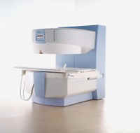
From Siemens Medical Systems;
This open MRI system is a dedicated extremity scanner, which is easy to use and easy to install. Whether you want to use it as a supplement to your whole-body scanner or you want to specialize in orthopedic exams,
MAGNETOM Jazz™ offers you a cost effective opportunity to make the most of your capabilities.
Device Information and Specification GRE, IR, FIR, STIR, TrueIR/FISP, FSE, MT, SS-FSE, MT-SE, MTC, MSE, GMR, fat/water sat./exc. IMAGING MODES Single, multislice, volume study, multi angle 512 x 512 full screen display POWER REQUIREMENTS 380/400/420/440/480 V | | | |
• View the DATABASE results for 'MAGNETOM Jazz™' (2).
| | | | |
| | | | | |
| |
|
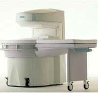
From Siemens Medical Systems;
the MAGNETOM Rhapsody™. This open MRI system offers the proven image
quality of 1.0 Tesla. In addition to the resulting broad range of applications, the open magnet of the high field system MAGNETOM Rhapsodyâ„¢ facilitates examination of claustrophobic and pediatric patients. And the system allows for expanded interventional applications.
Device Information and Specification CLINICAL APPLICATION Whole body GRE, IR, FIR, STIR, TrueIR/FISP, FSE, FLAIR, MT, SS-FSE, MT-SE, MTC, MSE, EPI, GMR, fat/water sat./exc. IMAGING MODES Single, multi slice, volume study, multi angle, multi oblique1024 x 1024 full screen display POWER REQUIREMENTS 380/400/420/440/480 V | | | |
• View the DATABASE results for 'MAGNETOM Rhapsody™' (2).
| | | | |
| | | Searchterm 'Slice' was also found in the following services: | | | | |
| | |
| |
|
( MRI) Magnetic resonance imaging is a noninvasive medical imaging technique that uses the interaction between radio frequency pulses, a strong magnetic field and body tissue to obtain images of slices/planes from inside the body. These magnets generate fields from approx. 2000 times up to 30000 times stronger than that of the Earth. The use of nuclear magnetic resonance principles produces extremely detailed pictures of the body tissue without the need for x-ray exposure and gives diagnostic information of various organs.
Measured are mobile hydrogen nuclei (protons are the hydrogen atoms of water, the 'H' in H 20), the majority of elements in the body. Only a small part of them contribute to the measured signal, caused by their different alignment in the magnetic field. Protons are capable of absorbing energy if exposed to short radio wave pulses (electromagnetic energy) at their resonance frequency. After the absorption of this energy, the nuclei release this energy so that they return to their initial state of equilibrium.
This transmission of energy by the nuclei as they return to their initial state is what is observed as the MRI signal. The subtle differing characteristic of that signal from different tissues combined with complex mathematical formulas analyzed on modern computers is what enables MRI imaging to distinguish between various organs. Any imaging plane, or slice, can be projected, and then stored or printed.
The measured signal intensity depends jointly on the spin density and the relaxation times ( T1 time and T2 time), with their relative importance depending on the particular imaging technique and choice of interpulse times. Any motion such as blood flow, respiration, etc. also affects the image brightness.
Magnetic resonance imaging is particularly sensitive in assessing anatomical structures, organs and soft tissues for the detection and diagnosis of a broad range of pathological conditions. MRI pictures can provide contrast between benign and pathological tissues and may be used to stage cancers as well as to evaluate the response to treatment of malignancies. The need for biopsy or exploratory surgery can be eliminated in some cases, and can result in earlier diagnosis of many diseases. See also MRI History and Functional Magnetic Resonance Imaging (fMRI). | | | | | |
• View the DATABASE results for 'Magnetic Resonance Imaging MRI' (9).
| | |
• View the NEWS results for 'Magnetic Resonance Imaging MRI' (222).
| | | | |  Further Reading: Further Reading: | | Basics:
|
|
News & More:
| |
| |
| | | Searchterm 'Slice' was also found in the following services: | | | | |
| | |
| |
|
On October 19, 2001, Philips Medical Systems completed an acquisition strategy through its purchase of Marconi Medical Systems.
The History of Marconi Medical Systems
2001
Royal Philips Electronics and Marconi plc announced that Philips has agreed to acquire Marconi Medical Systems for $1.1 billion.
2000
Marconi introduces Infinite Detector Technology for Mx8000 multi slice CT scanner, which acquires an unprecedented 16 simultaneous slices with sub-millimeter isotropic accuracy.
1999
At RSNA, Picker International unveils the new Marconi Medical Systems name and corporate vision.
1998
Picker International acquires the Computed Tomography Division of Elscint Ltd, immediately positioning Picker at the forefront of major global CT suppliers.
1986
Picker produces the industry's first 1.0T MR imager.
1981
Picker is sold to General Electric Co. Ltd. of England (GEC). Picker merged with Cambridge Instruments, GEC Medical, and American Optical to form Picker International.
1967
The name changed from Picker X-ray to Picker Corporation. Picker acquired Dunlee.
1946
The Dunlee Corporation started in Chicago by Dunmore Dunk and Zed. J. Atlee to meet demand for quality X-ray tubes and special purpose tubes.
1915
James Picker Company formed in New York City offering sales and service of X-ray equipment, film and accessories.
See also Philips Medical Systems and MRI History. | | | |
• View the DATABASE results for 'Marconi Medical Systems
' (3).
| | | | |
| | | | |
| | |
| | | |
|
| |
 | Look
Ups |
| |