 | Info
Sheets |
| | | | | | | | | | | | | | | | | | | | | | | | |
 | Out-
side |
| | | | |
|
| | | | | |  | Searchterm 'Slice' was also found in the following services: | | | | |
|  |  |
| |
|
The Dixon technique is a MRI method used for fat suppression and/or fat quantification. The difference in magnetic resonance frequencies between fat and water-bound protons allows the separation of water and fat images based on the chemical shift effect.
This imaging technique is named after Dixon, who published in 1984 the basic idea to use phase differences to calculate water and fat components in postprocessing. Dixon's method relies on acquiring an image when fat and water are 'in phase', and another in 'opposed phase' ( out of phase). These images are then added together to get water-only images, and subtracted to get fat-only images. Therefore, this sequence type can deliver up to 4 contrasts in one measurement: in phase, opposed phase, water and fat images. An additional benefit of Dixon imaging is that source images and fat images are also available to the diagnosing physician.
The original two point Dixon sequence (number of points means the number of images acquired at different TE) had limited possibilities to optimize the echo time, spatial resolution, slice thickness, and scan time; but Dixon based fat suppression can be very effective in areas of high magnetic susceptibility, where other techniques fail. This insensitivity to magnetic field inhomogeneity and the possibility of direct image-based water and fat quantification have currently generated high research interests and improvements to the basic method (three point Dixon).
The combination of Dixon with gradient echo sequences allows for example liver imaging with 4 image types in one breath hold. With Dixon TSE/FSE an excellent fat suppression with high resolution can be achieved, particularly useful in imaging of the extremities.
For low bandwidth imaging, chemical shift correction of fat images can be made before recombination with water images to produce images free of chemical shift displacement artifacts. The need to acquire more echoes lengthens the minimum scan time, but the lack of fat saturation pulses extends the maximum slice coverage resulting in comparable scan time. | | | | | |  Further Reading: Further Reading: | | Basics:
|
|
News & More:
| |
| |
| | | Searchterm 'Slice' was also found in the following services: | | | | |
| | |
| |
|
Device Information and Specification
CLINICAL APPLICATION
Whole body
SE, IR, FSE, FIR, GE, SG, BASG, PBSG, PCIR, DWI, Radial, Angiography: TOF, FLUTE (Fluoro-triggered bolus MRA), Time-resolved MRA
IMAGING MODES
Single, multislice, volume study
Level Range: -2,000 to +4,000
POWER REQUIREMENTS
208/220/240 V, single phase
| | | |
• View the DATABASE results for 'Echelon™ 1.5T' (2).
| | |
• View the NEWS results for 'Echelon™ 1.5T' (3).
| | | | | | Further Reading: | Basics:
|
|
| |
| | | | | |
| |
|
Motion of material being imaged, particularly flowing blood, can result in many possible effects in the images.
Fast moving blood produces flow voids,
blood flowing in to the outer slices of an imaging volume produces high signals ( flow related enhancement, entry slice phenomenon),
pulsatile flow creates ghost images of the vessel extending across the image in the phase encoding direction (image misregistration).
Flow-related dephasing occurring when spin isochromats are moving with different velocities in an external gradient field G so that they acquire different phases. When these phases vary by more then 180° within a voxel, substantial spin dephasing results leading to considerable intravascular signal loss.
These effects can be understood as caused by time of flight effects (washout or washin due to motion of nuclei between two consecutive spatially selective RF excitations, repeated in times on the order of, or shorter than the relaxation times of blood) or phase shifts (delay between phase encoding and frequency encoding) that can be acquired by excited spins moving along magnetic field gradients.
The inconsistency of the signal resulting from pulsatile flow can lead to artifacts in the image. The flow effects can also be exploited for MR angiography or flow measurements.
See also Flow Artifact. | | | | | |
• View the DATABASE results for 'Flow Effects' (16).
| | | | | | Further Reading: | News & More:
|
|
| |
| | | Searchterm 'Slice' was also found in the following services: | | | | |
| | |
| |
|
(HS) A method in which approximately one half of the acquisition matrix in the phase encoding direction is acquired. Half scan is possible because of symmetry in acquired data. Since negative values of phase encoded measurements are identical to corresponding positive values, only a little over half (more than 62.5%) of a scan actually needs to be acquired to replicate an entire scan.
This results in a reduction in scan time at the expense of signal to noise ratio. The time reduction can be nearly a factor of two, but full resolution is maintained.
Half scan can be used when scan times are long, the signal to noise ratio is not critical and where full spatial resolution is required. Half scan is particularly appropriate for scans with a large field of view and relatively thick slices; and, in 3D scans with many slices.
In some fast scanning techniques the use of Half scan enables a shorter TE thus improving contrast. For this reason, the Half scan parameter is located in the contrast menu.
More information about scan time reduction; see also partial fourier technique. | | | |
• View the DATABASE results for 'Half Scan' (4).
| | | | |
| | | Searchterm 'Slice' was also found in the following services: | | | | |
| | |
| |
|
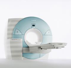
From Siemens Medical Systems;
MAGNETOM Avanto with Tim - Total imaging matrix technology.
For true whole-body anatomical coverage. For ultra-fast image
acquisition. Aids the physician in fast and precise
evaluation of systemic diseases like colon cancer, metastasis imaging, vessel diseases, and preventional exams. For claustrophobic patients,
MAGNETOM Avanto enables feetfirst exams for nearly all MR procedures. For obese patients, MAGNETOM Avanto supports up to 200 kg (400 lbs), without table movement restrictions. The AudioComfort technology enables up to a 30 dB(A) acoustic noise reduction, that means nearly all clinical routine sequences are running under 99 dB(A).
Device Information and Specification
CLINICAL APPLICATION
Whole body
CONFIGURATION
Compact short bore
Body, Tim [32 x 8], Tim [76 x18], Tim [76 coil elements with up to 32 RF channels]
GRE, IR, FIR, STIR, TrueIR/FISP, FSE, FLAIR, MT, SS-FSE, MT-SE, MTC, MSE, EPI, 3D DESS//CISS/PSIF, GMR
IMAGING MODES
Single, multi slice, volume study, multi angle, multi oblique
1024 x 1024 full screen display
POWER REQUIREMENTS
380/400/420/440/480 V
Passive, act.; 1st order std./2nd opt.
| | | |
• View the DATABASE results for 'MAGNETOM Avanto™' (2).
| | | | | | Further Reading: | Basics:
|
|
News & More:
| |
| |
| | | | |
| | | |
|
| |
 | Look
Ups |
| |


