 | Info
Sheets |
| | | | | | | | | | | | | | | | | | | | | | | | |
 | Out-
side |
| | | | |
|
| | | | |
Result : Searchterm 'Signal to Noise Ratio' found in 1 term [ ] and 49 definitions [ ] and 49 definitions [ ] ]
| | previous 41 - 45 (of 50) nextResult Pages : [1] [2 3 4 5 6 7 8 9 10] |  | |  | Searchterm 'Signal to Noise Ratio' was also found in the following services: | | | | |
| |  |
| |
|
| | | | | |  Further Reading: Further Reading: | News & More:
|
|
| |
| | | Searchterm 'Signal to Noise Ratio' was also found in the following services: | | | | |
| | |
| |
|
Liver imaging with gadolinium contrast enhanced MRI is sometimes not sufficient for a reliable diagnosis of liver lesions.
For this reasons, special liver Contrast agents that are targeted to the reticuloendothelial system (RES), have been developed to improve both detection and characterization of liver and spleen lesions. Reticuloendothelial Contrast Agents, as e.g. superparamagnetic iron oxides ( SPIO), are taken up by healthy liver tissue but not tumors.
These RES targeted contrast agents provide a prolonged imaging window and enough time for high spatial resolution or multiple breath hold images. Reticuloendothelial contrast agents have an increased sensitivity for the detection of small liver lesions (e.g., metastases), compared with gadolinium enhanced MRI and spiral CT. At higher field strengths with an increased signal to noise ratio the susceptibility effect with iron oxide particles may be enhanced.
Other new agents ( Gadobenate Dimeglumine, Gadoxetic Acid) have both an initial extracellular circulation and a delayed liver-specific uptake. Since a considerable part of these contrast agents is excreted in the bile, functional biliary imaging can diagnose biliary anomalies, postoperative bile leaks, and anastomotic strictures. Other agents, such as liposomes (with encapsulated Gd-DTPA) or DOTA complexes are in different development stages.
See also Hepatobiliary Contrast Agents, Gadolinium Oxide, Superparamagnetic Iron Oxide and Liposomes. | | | |
• View the DATABASE results for 'Reticuloendothelial Contrast Agents' (3).
| | | | |
| | | | | |
| |
|
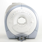
From GE Healthcare;
The GE Signa HDx MRI system is a whole body magnetic resonance scanner designed to support high resolution, high signal to noise ratio, and short scan times.
The 1.5T Signa HDx MR Systems is a modification of the currently marketed GE 1.5T machines, with the main difference being the change to the receive chain architecture that includes a thirty two independent receive channels, and allows for future expansion in 16 channel increments. The overall system has been improved with a simplified user interface
and a single 23" liquid crystal display, improved multi channel surface coil connectivity, and an improved image reconstruction architecture known as the Volume Recon Engine (VRE).
Device Information and Specification CLINICAL APPLICATION Whole body CONFIGURATION Compact short bore Standard: SE, IR, 2D/3D GRE and SPGR, Angiography: 2D/3D TOF, 2D/3D Phase Contrast; 2D/3D FSE, 2D/3D FGRE and FSPGR, SSFP, FLAIR, EPI, optional: 2D/3D Fiesta, FGRET, Spiral, Tensor, 2D 0.7 mm to 20 mm; 3D 0.1 mm to 5 mm 128x512 steps 32 phase encode POWER REQUIREMENTS 480 or 380/415 less than 0.03 L/hr liquid helium | | | | | |
| | | Searchterm 'Signal to Noise Ratio' was also found in the following services: | | | | |
| | |
| |
|

From GE Healthcare;
The Signa HDx MRI system is GE's leading edge whole body magnetic resonance scanner designed to support high resolution, high signal to noise ratio, and short scan times.
Signa HDx 3.0T offers new technologies like ultra-fast image reconstruction through the new XVRE recon engine, advancements in parallel imaging algorithms and the broadest range of premium applications. The HD applications, PROPELLER (high-quality brain imaging extremely resistant to motion artifacts), TRICKS (contrast-enhanced angiographic vascular lower leg imaging), VIBRANT (for breast MRI), LAVA (high resolution liver imaging with shorter breath holds and better organ coverage) and MR Echo (high-definition cardiac images in real time) offer unique capabilities.
Device Information and Specification CLINICAL APPLICATION Whole body
CONFIGURATION Compact short bore SE, IR, 2D/3D GRE, RF-spoiled GRE, 2DFGRE, 2DFSPGR, 3DFGRE, 3DFSPGR, 3DTOFGRE, 3DFSPGR, 2DFSE, 2DFSE-XL, 2DFSE-IR, T1-FLAIR, SSFSE, EPI, DW-EPI, BRAVO, Angiography: 2D/3D TOF, 2D/3D phase contrast vascular IMAGING MODES Single, multislice, volume study, fast scan, multi slab, cine, localizer H*W*D 240 x 2216,6 x 201,6 cm POWER REQUIREMENTS 480 or 380/415, 3 phase ||
COOLING SYSTEM TYPE Closed-loop water-cooled grad. | | | | | |
| | | Searchterm 'Signal to Noise Ratio' was also found in the following services: | | | | |
| | |
| |
|
| | | |
• View the DATABASE results for 'Signal Averaging' (6).
| | | | |
| | | | |
| | |
| | | |
|
| |
 | Look
Ups |
| |


