Magnetic Resonance - Technology Information Portal Welcome to MRI Technology
Info
  Sheets

Out-
      side


 'Resolution' 
SEARCH FOR    
 
  2 3 5 A B C D E F G H I J K L M N O P Q R S T U V W X Y Z
Result : Searchterm 'Resolution' found in 8 terms [] and 144 definitions []
previous     36 - 40 (of 152)     next
Result Pages : [1 2]  [3 4 5 6 7 8 9 10 11 12 13 14 15 16 17 18 19 20 ... ]
Searchterm 'Resolution' was also found in the following services: 
spacer
News  (40)  Resources  (13)  Forum  (15)  
 
MS-325Forum -
related threadsInfoSheet: - Contrast Agents - 
Intro, Overview, 
Characteristics, 
Types of, 
etc.MRI Resource Directory:
 - Contrast Agents -
 
MS-325 is the formerly code name of gadofosveset trisodium (new trade name Vasovist). MS-325 belongs to a new class of blood pool agents for magnetic resonance angiography (MRA) to diagnose vascular disease. Gadofosveset trisodium has ten times the signal-enhancing power of existing contrast agents as well as prolonged retention in the blood. This enables the rapid acquisition of high resolution MRA's using standard MRI machines.
Gadofosveset trisodium, which is gadolinium-based, stays in the blood stream as a result of transient binding to albumin. Albumin binding offers an additional benefit beyond localization in the blood pool. The contrast agent begins to spin much more slowly, at the rate albumin spins, causing a relaxivity gain that produces a substantially brighter signal than would be possible with freely circulating gadolinium. MS-325 is an intravascular contrast agent intended for use in MRI as an aid in diagnosing aortoiliac occlusive disease in patients with known or suspected peripheral vascular disease (PVD) or abdominal aortic aneurysm (AAA).
Currently clinical trials completed for peripheral vascular disease and coronary artery disease. Additional trials are also being conducted to evaluate MS-325 as an aid in diagnosing breast cancer and suggested that it might be feasible to combine the use of MS-325, injected during peak stress, with delayed high-resolution imaging to identify myocardial perfusion defects.
Vasovist (MS-325) would compete with the contrast agents Ferumoxytol (Code 7228) from AMAG Pharmaceuticals, Inc. and NC100150 Injection from Nycomed Amersham, but their further development is uncertain.
Partners in development: EPIX Pharmaceuticals, Inc., Mallinckrodt Inc., and Bayer Schering Pharma AG. Bayer Schering Pharma has the worldwide marketing rights for the product.
Formerly known under the Mallinckrodt trademark name, AngioMARK®.

See also Classifications, Characteristics, etc.
spacer

• View the NEWS results for 'MS-325' (10).Open this link in a new window.
 
Further Reading:
  News & More:
EPIX Medical's new multimedia Web site features AngioMARK images in 3D
Friday, 5 March 1999
MRI technology combined with contrast agent optimizes diagnosis of cardiovascular disease
1999
Searchterm 'Resolution' was also found in the following services: 
spacer
Radiology  (27) Open this link in a new windowUltrasound  (69) Open this link in a new window
MSK-Extremeâ„¢InfoSheet: - Devices -
Intro, 
Types of Magnets, 
Overview, 
etc.MRI Resource Directory:
 - Devices -
 
www.onicorp.com/ From ONI Medical Systems, Inc.;
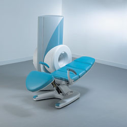
MSK-Extremeâ„¢MRI system is a dedicated high field extremity imaging device, designed to provide orthopedic surgeons and other physicians with detailed diagnostic images of the foot, ankle, knee, hand, wrist and elbow, all with the clinical confidence and advantages derived from high field, whole body MRI units. The light weight (less than 650 kg) of the OrthOne System performs rapid patient studies, is easy to operate, has a patient friendly open environment and can be installed in a practice office or hospital, all at a cost similar to a low field extremity machine.
New features include a more powerful operating system that offers increased scan speed as well as a 160-mm knee coil with higher signal to noise ratio, and the option of a CD burner.
Device Information and Specification
CLINICAL APPLICATION
Dedicated extremity imaging
CONFIGURATION
16 cm knee, 18 cm lower extremity;; 12.3 cm upper extremity, additional high resolution v-SPEC Coils: 80 mm, 100 mm, or 145 mm.
SYNCHRONIZATION
No
PULSE SEQUENCES
SE, FSE, GE2D, GE3D, Inversion recovery (IR), Driven Equilibrium, Fat Saturation (FS), STIR, MT, PD, Flow Compensation (FC), RF spoiling, MTE, No Phase Wrap (NPW)
IMAGING MODES
Scout, single, multislice, volume
TR
10-10,000ms; 1ms steps
TE
5-150ms; 1 ms steps
SINGLE/MULTI SLICE
2D less than 200 msec/image
4cm-16cm
2D: 2mm-10mm/.1mm incr.
Up to 1,000x1,000
MEASURING MATRIX
X/Y: 64-512; 2 pixel steps
PIXEL INTENSITY
4,096 grey lvls; 256 lvls in 3D
28cm ID x 50cm L
MAGNET WEIGHT
635 kg
H*W*D
146 x 69 x 84 cm
POWER REQUIREMENTS
115VAC, 1phase, 20A; 208VAC, 3 phase, 30A
COOLING SYSTEM TYPE
LHe with 2 stage cold head
Negligible
STRENGTH
15 mT/m
5-GAUSS FRINGE FIELD
1.25m radial x 1.8m axial
Passive
spacer
 
Further Reading:
  Basics:
MSK Extreme Brochure(.pdf)
   by www.nova-logic.ch    
MSK Extreme Specifications(.pdf)
   by www.nova-logic.ch    
MRI Resources 
Education pool - MRI Accidents - Manufacturers - Stimulator pool - Spectroscopy - Contrast Enhanced MRI
 
Multiple Overlapping Thin Slab (Slice) Acquisition
 
(MOTSA) This technique combines the best features of 2D time of flight angiography (2D TOF) and 3D TOF MRA. The MOTSA technique consists of multiple 2 cm thick 3D TOF slabs (which minimize saturation effects for through plane flow) combine to provide unlimited coverage similar to multiple 2D TOF slices. High resolution imaging of the carotid arteries is possible when image quality is of greater concern than acquisition time. Images with 1 mm (or less) spatial resolution in all three planes are required. The slabs typically overlap 25-40 to minimize the venetian blind artifact venetian blind artifact due to minimal saturation effects. MOTSA is an useful technique for the evaluation of vertebrobasilar ischemia and aneurysm scanning from the foramen magnum through the circle of Willis.
 
Images, Movies, Sliders:
 Circle of Willis, Time of Flight, MIP  Open this link in a new window
    
SlidersSliders Overview

 
spacer
Searchterm 'Resolution' was also found in the following services: 
spacer
News  (40)  Resources  (13)  Forum  (15)  
 
Partial Averaging
 
Partial averaging is a scan time reduction method that takes advantage of the complex conjugate of the k-space. The number of phase encoding steps of the acquisition matrix are reduced in the phase encoding direction.
Since negative values of phase encoded measurements are identical to corresponding positive values, only a little over half (more than 62.5%) of a scan actually needs to be acquired to replicate an entire scan. This results in a reduction in scan time at the expense of signal to noise ratio. The time reduction can be nearly a factor of two, but full resolution is maintained.
Partial Fourier averaging can be used when scan times are long, the signal to noise ratio is not critical and where full spatial resolution is required. Partial averaging is particularly appropriate for scans with a large field of view and relatively thick slices; and in 3D scans with many slices. In some fast scanning techniques the use of partial averaging enables a shorter TE thus improving contrast.
Partial averaging is also called Fractional NEX, Half Scan, Half Fourier, Phase Conjugate Symmetry, Single Side Encoding.
spacer

• View the DATABASE results for 'Partial Averaging' (4).Open this link in a new window

Searchterm 'Resolution' was also found in the following services: 
spacer
Radiology  (27) Open this link in a new windowUltrasound  (69) Open this link in a new window
Partial Fourier Imaging
 
Reconstruction of an image from a MR data set comprising an asymmetric sampling of k-space. For example, it can be used either to shorten image acquisition time, by reducing the number of phase encoding steps required, or to shorten the echo time, TE, by moving the echo off-center in the acquisition window. In either case the signal to noise ratio is reduced and the resolution can be improved to correspond to the maximum available resolution in the data.
spacer

• View the DATABASE results for 'Partial Fourier Imaging' (5).Open this link in a new window

MRI Resources 
Contrast Enhanced MRI - Journals - Service and Support - MRCP - MR Myelography - Health
 
previous      36 - 40 (of 152)     next
Result Pages : [1 2]  [3 4 5 6 7 8 9 10 11 12 13 14 15 16 17 18 19 20 ... ]
 Random Page
 
Share This Page
FacebookTwitterLinkedIn

MR-TIP    
Community   
User
Pass
Forgot your UserID/Password ?    


How AI will impact MRI :
only diagnostics 
saving time 
reducing cost 
makes planning obsolete 
reduce human knowledge 
not at all 

Look
      Ups


MR-TIP.com uses cookies! By browsing MR-TIP.com, you agree to our use of cookies.

Magnetic Resonance - Technology Information Portal
Member of SoftWays' Medical Imaging Group - MR-TIP • Radiology-TIP • Medical-Ultrasound-Imaging • 
Copyright © 2003 - 2024 SoftWays. All rights reserved. [ 24 April 2024]
Terms of Use | Privacy Policy | Advertising
 [last update: 2024-02-26 03:41:00]