| Info
Sheets |
| | | | | | | | | | | | | | | | | | | | | | | | |
 | Out-
side |
| | | | |
|
| | | | | |  | Searchterm 'Resolution' was also found in the following services: | | | | |
|  |  |
| |
|
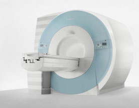
Device Information and Specification
CLINICAL APPLICATION
Whole body
CONFIGURATION
Short bore compact
Standard: Head, body, cardiac, optional phased array: Spine, pediatric, 3rd party connector; Optional SENSE? coils for all applications
SE, Modified-SE, IR (T1, T2, PD), STIR, FLAIR, SPIR, FFE, T1-FFE, T2-FFE, Balanced FFE, TFE, Balanced TFE, Dynamic, Keyhole, 3D, Multi Chunk 3D, Multi Stack 3D, K Space Shutter, MTC, TSE, Dual IR, DRIVE, EPI, Cine, 2DMSS, DAVE, Mixed Mode; Angiography: Inflow MRA, TONE, PCA, CE MRA
128 x 128, 256 x 256,512 x 512,1024 x 1024 (64 for Bold img)
Variable in 1% increments
Lum.: 120 cd/m2; contrast: 150:1
Variable (op. param. depend.)
POWER REQUIREMENTS
380/400 V
| | | | | |  Further Reading: Further Reading: | News & More:
|
|
| |
| | | Searchterm 'Resolution' was also found in the following services: | | | | |
| | |
| |
|
Knee MRI, with its high soft tissue contrast is one of the main imaging tools to depict knee joint pathology. MRI allows accurate imaging of intra-articular structures such as ligaments, cartilage, menisci, bone marrow, synovium, and adjacent soft tissue.
Knee exams require a dedicated extremity coil, providing a homogenous imaging volume and high SNR to ensure best signal coverage.
A complete knee MR examination includes for example sagittal and coronal T1 weighted, and proton density weighted pulse sequences +/- fat saturation, or STIR sequences. For high spatial resolution, maximal 4 mm thick slices with at least an in plane resolution of 0.75 mm and small gap are recommended. To depict the anterior cruciate ligament clearly, the sagittal plane has to be rotated 10 - 20° externally (parallel to the medial border of the femoral condyle). Retropatellar cartilage can bee seen for example in axial T2 weighted gradient echo sequences with Fatsat. However, the choice of the pulse sequences is depended of the diagnostic question, the used scanner, and preference of the operator.
Diagnostic quality in knee imaging is possible with field strengths ranging from 0.2 to 3T. With low field strengths more signal averages must be measured, resulting in increased scan times to provide equivalent quality as high field strengths.
More diagnostic information of meniscal tears and chondral defects can be obtained by direct magnetic resonance arthrography, which is done by introducing a dilute solution of gadolinium in saline (1:1000) into the joint capsule. The knee is then scanned in all three planes using T1W sequences with fat suppression. For indirect arthrography, the contrast is given i.v. and similar scans are started 20 min. after injection and exercise of the knee.
Frequent indications of MRI scans in musculoskeletal knee diseases are: e.g., meniscal degeneration and tears, ligament injuries, osteochondral fractures, osteochondritis dissecans, avascular bone necrosis and rheumatoid arthritis. See also Imaging of the Extremities and STIR. | | | | | |
• View the DATABASE results for 'Knee MRI' (4).
| | |
• View the NEWS results for 'Knee MRI' (4).
| | | | | | Further Reading: | | Basics:
|
|
News & More:
| |
| |
| | | | | |
| |
|
Liver imaging can be performed with sonography, computed tomography (CT) and magnetic resonance imaging ( MRI). Ultrasound is, caused by the easy access, still the first-line imaging method of choice; CT and MRI are applied whenever ultrasound imaging yields vague results. Indications are the characterization of metastases and primary liver tumors e.g., benign lesions such as focal nodular hyperplasia (FNH), adenoma, hemangioma and malignant lesions (cancer) such as hepatocellular carcinomas (HCC).
The decision, which medical imaging modality is more suitable, MRI or CT, is dependent on the different factors. CT is less costly and more widely available; modern multislice scanners provide high spatial resolution and short scan times but has the disadvantage of radiation exposure.
With the introduction of high performance MR systems and advanced sequences the image quality of MRI for the liver has gained substantially. Fast spin echo or single shot techniques, often combined with fat suppression, are the most common T2 weighted sequences used in liver MRI procedures.
Spoiled gradient echo sequences are used as ideal T1 weighted sequences for evaluating of the liver. The repetition time (TR) can be sufficiently long to acquire enough sections covering the entire liver in one pass, and to provide good signal to noise. The TE should be the shortest in phase echo time (TE), which provides strong T1 weighting, minimizes magnetic susceptibility effects, and permits acquisition within one breath hold to cover the whole liver. A flip angle of 80° provides good T1 weighting and less of power deposition and tissue saturation than a larger flip angle that would provide comparable T1 weighting.
Liver MRI is very dependent on the administration of contrast agents, especially when detection and characterization of focal lesions are the issues. Liver MRI combined with MRCP is useful to evaluate patients with hepatic and biliary disease.
Gadolinium chelates are typical non-specific extracellular agents diffusing rapidly to the extravascular space of tissues being cleared by glomerular filtration at the kidney. These characteristics are somewhat problematic when a large organ with a huge interstitial space like the liver is imaged. These agents provide a small temporal imaging window (seconds), after which they begin to diffuse to the interstitial space not only of healthy liver cells but also of lesions, reducing the contrast gradient necessary for easy lesion detection. Dynamic MRI with multiple phases after i.v. contrast media (Gd chelates), with arterial, portal and late phase images (similar to CT) provides additional information.
An additional advantage of MRI is the availability of liver-specific contrast agents (see also Hepatobiliary Contrast Agents). Gd-EOB-DTPA (gadoxetate disodium, Gadolinium ethoxybenzyl dimeglumine, EOVIST Injection, brand name in other countries is Primovist) is a gadolinium-based MRI contrast agent approved by the FDA for the detection and characterization of known or suspected focal liver lesions.
Gd-EOB-DTPA provides dynamic phases after intravenous injection, similarly to non-specific gadolinium chelates, and distributes into the hepatocytes and bile ducts during the hepatobiliary phase. It has up to 50% hepatobiliary excretion in the normal liver.
Since ferumoxides are not eliminated by the kidney, they possess long plasmatic half-lives, allowing circulation for several minutes in the vascular space. The uptake process is dependent on the total size of the particle being quicker for larger particles with a size of the range of 150 nm (called superparamagnetic iron oxide). The smaller ones, possessing a total particle size in the order of 30 nm, are called ultrasmall superparamagnetic iron oxide particles and they suffer a slower uptake by RES cells. Intracellular contrast agents used in liver MRI are primarily targeted to the normal liver parenchyma and not to pathological cells. Currently, iron oxide based MRI contrast agents are not marketed.
Beyond contrast enhanced MRI, the detection of fatty liver disease and iron overload has clinical significance due to the potential for evolution into cirrhosis and hepatocellular carcinoma. Imaging-based liver fat quantification (see also Dixon) provides noninvasively information about fat metabolism; chemical shift imaging or T2*-weighted imaging allow the quantification of hepatic iron concentration.
See also Abdominal Imaging, Primovistâ„¢, Liver Acquisition with Volume Acquisition (LAVA), T1W High Resolution Isotropic Volume Examination (THRIVE) and Bolus Injection.
For Ultrasound Imaging (USI) see Liver Sonography at Medical-Ultrasound-Imaging.com. | | | | | | | | | | |
• View the DATABASE results for 'Liver Imaging' (13).
| | |
• View the NEWS results for 'Liver Imaging' (10).
| | | | | | Further Reading: | | Basics:
|
|
News & More:
|  |
Utility and impact of magnetic resonance elastography in the clinical course and management of chronic liver disease
Saturday, 20 January 2024 by www.nature.com | | |
Even early forms of liver disease affect heart health, Cedars-Sinai study finds
Thursday, 8 December 2022 by www.eurekalert.org | | |
For monitoring purposes, AI-aided MRI does what liver biopsy does with less risk, lower cost
Wednesday, 28 September 2022 by radiologybusiness.com | | |
Perspectum: High Liver Fat (Hepatic Steatosis) Linked to Increased Risk of Hospitalization in COVID-19 Patients With Obesity
Monday, 29 March 2021 by www.businesswire.com | | |
EMA's final opinion confirms restrictions on use of linear gadolinium agents in body scans
Friday, 21 July 2017 by www.ema.europa.eu | | |
T2-Weighted Liver MRI Using the MultiVane Technique at 3T: Comparison with Conventional T2-Weighted MRI
Friday, 16 October 2015 by www.ncbi.nlm.nih.gov | | |
EORTC study aims to qualify ADC as predictive imaging biomarker in preoperative regimens
Monday, 4 January 2016 by www.eurekalert.org | | |
MRI effectively measures hemochromatosis iron burden
Saturday, 3 October 2015 by medicalxpress.com | | |
Total body iron balance: Liver MRI better than biopsy
Sunday, 15 March 2015 by www.eurekalert.org |
|
| |
| | | Searchterm 'Resolution' was also found in the following services: | | | | |
| | |
| |
|
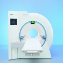
Device Information and Specification
CLINICAL APPLICATION
Whole body
GRE, IR, FIR, STIR, TrueIR/FISP, FSE, FLAIR, MT, SS-FSE, MT-SE, MTC, MSE, EPI, GMR, fat/water sat./exc.
IMAGING MODES
Single, multislice, volume study, multi angle, multi oblique
TR
2.4 msec std.; 2.0 opt.; 1.8 w/ 30mT/m at 256matrix
TE
1.1 msec std.; 0.9 opt.; 0.78 w/30 mT/m at 256matrix
178 images/sec at 256 x 256 at 100% FOV
1024 x 1024 full screen display
21 micrometer in plane, 11 micrometer optional
MAGNET WEIGHT (gantry included)
3500kg, 5000kg in operation
DIMENSION H*W*D (gantry included)
POWER REQUIREMENTS
380/400/420/440/480 V
STRENGTH
20/35 mT/m standard, 30/52 opt.
Passive, act.; 1st order std./2nd opt.
| | | |
• View the DATABASE results for 'MAGNETOM Harmony™' (2).
| | | | | | Further Reading: | Basics:
|
|
| |
| | | Searchterm 'Resolution' was also found in the following services: | | | | |
| | |
| |
|
Device Information and Specification CLINICAL APPLICATION Whole body SE, IR, 2D/3D TurboSE, Turbo IR, Dark-Fluid IR, True IR, 2D/3D MEDIC, 2D/3D GRE FLASH, 2D/3D GRE FISP, 2D/3D PSIF, 2D TurboFLASH, 3D MP-RAGE, 3D TurboFLASH, 2D/3D TOF angiography, MTC, TONE with 3D TOF MRA, GMR, LOTA IMAGING MODES Single, multislice, volume study, multi angle, multi oblique178 images/sec at 256 x 256 at 100% FOV1024 x 1024 full screen display POWER REQUIREMENTS 380/400/420/440/480 V Passive, act.; 1st order std./2nd opt. | | | | | |
| | | | |
| | | |
|
| |
 | Look
Ups |
| |