 | Info
Sheets |
| | | | | | | | | | | | | | | | | | | | | | | | |
 | Out-
side |
| | | | |
|
| | | | |
Result : Searchterm 'Open MRI' found in 1 term [ ] and 36 definitions [ ] and 36 definitions [ ] ]
| | previous 31 - 35 (of 37) nextResult Pages : [1] [2 3 4 5 6 7 8] |  | |  | Searchterm 'Open MRI' was also found in the following services: | | | | |
| |  |
| |
|
From MagneVu;
The MagneVu 1000 is a compact, robust, and portable, permanent magnet MRI system and operates without special shielding or costly site preparation.
This MRI device utilizes a patented non-homogeneous magnetic field image acquisition method to achieve high performance imaging. The MagneVu 1000 MRI scanner is designed for MRI of the extremities with the current specialty areas in diabetes and rheumatoid arthritis. Easy access is afforded for claustrophobic, pediatric, or limited mobility patients. In August 1998
FDA marketing clearance and other regulatory approvals have been received. Until 2008, over 130 devices in the US are in use. Some further developments of MagneVu's extremity scanner are: 'truly Plug n' Play MRI™' and iSiS ( which adds wireless capability to the second generation MV1000-XL).
Device Information and Specification IMAGING MODES 3-dimensional multi-echo data acquisition | | | | | |  Further Reading: Further Reading: | News & More:
|
|
| |
| | | Searchterm 'Open MRI' was also found in the following services: | | | | |
| | |
| |
|
The definition of imaging is the visual representation of an object. Medical imaging began after the discovery of x-rays by Konrad Roentgen 1896. The first fifty years of radiological imaging, pictures have been created by focusing x-rays on the examined body part and direct depiction onto a single piece of film inside a special cassette. The next development involved the use of fluorescent screens and special glasses to see x-ray images in real time.
A major development was the application of contrast agents for a better image contrast and organ visualization. In the 1950s, first nuclear medicine studies showed the up-take of very low-level radioactive chemicals in organs, using special gamma cameras. This medical imaging technology allows information of biologic processes in vivo. Today, PET and SPECT play an important role in both clinical research and diagnosis of biochemical and physiologic processes. In 1955, the first x-ray image intensifier allowed the pick up and display of x-ray movies.
In the 1960s, the principals of sonar were applied to diagnostic imaging. Ultrasonic waves generated by a quartz crystal are reflected at the interfaces between different tissues, received by the ultrasound machine, and turned into pictures with the use of computers and reconstruction software. Ultrasound imaging is an important diagnostic tool, and there are great opportunities for its further development. Looking into the
future, the grand challenges include targeted contrast agents, real-time 3D ultrasound imaging, and molecular imaging.
Digital imaging techniques were implemented in the 1970s into conventional fluoroscopic image intensifier and by Godfrey Hounsfield with the first computed tomography. Digital images are electronic snapshots sampled and mapped as a grid of dots or pixels. The introduction of x-ray CT revolutionised medical imaging with cross sectional images of the human body and high contrast between different types of soft tissue. These developments were made possible by analog to digital converters and computers. The multislice spiral CT technology has expands the clinical applications dramatically.
The first MRI devices were tested on clinical patients in 1980. The spread of CT machines is the spur to the rapid development of MRI imaging and the introduction of tomographic imaging techniques into diagnostic nuclear medicine. With technological improvements including higher field strength, more open MRI magnets, faster gradient systems, and novel data-acquisition techniques, MRI is a real-time interactive imaging modality that provides both detailed structural and functional information of the body.
Today, imaging in medicine has advanced to a stage that was inconceivable 100 years ago, with growing medical imaging modalities:
•
Single photon emission computed tomography (SPECT)
•
Positron emission tomography (PET)
All this type of scans are an integral part of modern healthcare.
Because of the rapid development of digital imaging modalities, the increasing need for an efficient management leads to the widening of radiology information systems (RIS) and archival of images in digital form in picture archiving and communication systems (PACS).
In telemedicine, healthcare professionals are linked over a computer network. Using cutting-edge computing and communications technologies, in videoconferences, where audio and visual images are transmitted in real time, medical images of MRI scans, x-ray examinations, CT scans and other pictures are shareable.
See also Hybrid Imaging.
See also the related poll results: ' In 2010 your scanner will probably work with a field strength of', ' MRI will have replaced 50% of x-ray exams by' | | | | | | | | |
• View the DATABASE results for 'Medical Imaging' (20).
| | |
• View the NEWS results for 'Medical Imaging' (81).
| | | | | | Further Reading: | | Basics:
|
|
News & More:
| |
| |
| | | | | |
| |
|
From Odin Medical Technologies, Inc.;
the PoleStar™ N-10 is a compact, mobile MRI scanner that mounts to a standard operating room table. The magnets raise into position for imaging, but lower to make surgery easier, and the low magnetic field makes it possible to use many conventional surgical instruments.
When not in use, the PoleStar™ is stored in a nearby closet that allows the room to be used for conventional surgical procedures. The PoleStar™ N-10 is supplied with a fully integrated image guidance system that utilizes intraoperatively acquired images.
The successor, the new PoleStar™ N20 sets a new standard in intraoperative magnetic resonance imaging.
Device Information and Specification CLINICAL APPLICATION Intraoperative | | | |
• View the DATABASE results for 'PoleStar™' (2).
| | | | |
| | | Searchterm 'Open MRI' was also found in the following services: | | | | |
| | |
| |
|
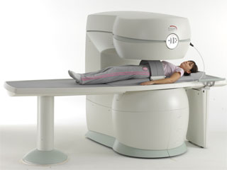
From Esaote S.p.A.;
Esaote introduced the S-SCAN at RSNA in November 2007. The S-SCAN is a dedicated joint and spine MR scanner derived from the company's earlier G-SCAN system. Unlike the G-SCAN, neither the patient table nor
the magnet can rotate from horizontal to vertical position. The patient table can only moved manually. Improved electronics, new coils for lumbar and cervical spine, new pulse sequences, a modified version of the magnet poles and gradient coils are used with a new software release in the S-SCAN.
Esaote North America is the exclusive U.S. distributor of this MRI device.
Device Information and Specification SE, GE, IR, STIR, TSE, 3D CE, GE-STIR, 3D GE, ME, TME, HSE POWER REQUIREMENTS 3 kW; 110/220 V single phase | | | |
• View the DATABASE results for 'S-SCAN' (3).
| | |
• View the NEWS results for 'S-SCAN' (1).
| | | | | | Further Reading: | News & More:
|
|
| |
| | | Searchterm 'Open MRI' was also found in the following services: | | | | |
| | |
| |
|

From GE Healthcare;
The Signa SP 0.5T™ is an open MRI magnet that is designed for use in interventional radiology and intra-operative imaging. The vertical gap configuration increases patient positioning options, improves patient observation, and allows continuous access to the patient during imaging.
The magnet enclosure also incorporates an intercom, patient observation video camera, laser patient alignment lights, and task lighting in the imaging volume.
Device Information and Specification CLINICAL APPLICATION Whole body Integrated transmit and receive body coil; optional rotational body coil, head; other coils optional; open architecture makes system compatible with a wide selection of coilsarray Standard: SE, IR, 2D/3D GRE and SPGR, 2D/3D TOF, 2D/3D FSE, 2D/3D FGRE and FSPGR, SSFP, FLAIR, EPI, optional: 2D/3D Fiesta, true chem sat, fat/water separation, single shot diffusion EPI IMAGING MODES Localizer, single slice, multislice, volume, fast, POMP, multi slab, cine, slice and frequency zip, extended dynamic range, tailored RF TR 1.3 to 12000 msec in increments of 1 msec TE 0.4 to 2000 msec in increments of 1 msec 2D: 1.4mm - 20mm 3D: 0.2mm - 20mm POWER REQUIREMENTS 200 - 480, 3-phase | | | |
• View the DATABASE results for 'Signa SP 0.5T™ Open Configuration' (2).
| | | | | | Further Reading: | News & More:
|
|
| |
| | | | |
| | |
| | | |
|
| |
 | Look
Ups |
| |


