 | Info
Sheets |
| | | | | | | | | | | | | | | | | | | | | | | | |
 | Out-
side |
| | | | |
|
| | | | |
Result : Searchterm 'Open MRI' found in 1 term [ ] and 36 definitions [ ] and 36 definitions [ ] ]
| | previous 26 - 30 (of 37) nextResult Pages : [1] [2 3 4 5 6 7 8] |  | |  | Searchterm 'Open MRI' was also found in the following services: | | | | |
| |  |
| |
|
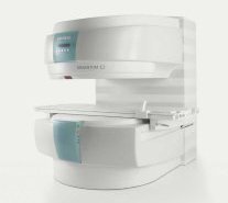
From Siemens Medical Systems;
A new, powerful, compact player in MRI. For both, patients and health care professionals, the mid-field has realized a giant step to cost efficient quality care. Obese patients and people with claustrophobia appreciate the comfortable side loading. The smallest pole diameter - 137 cm (54 inches) allows for optimal patient comfort.
Device Information and Specification
CLINICAL APPLICATION
Whole body
SE, FLASH, FISP, IR, FIR, STIR, TrueIR/FISP, FSE, MT, SS-FSE, MT-SE, MTC, MSE, EPI, PSIF
IMAGING MODES
Single, multislice, volume study, multi angle, multi oblique
512 x 512 full screen display
41 cm vertical gap distance
| | | | | | | | | | |  Further Reading: Further Reading: | Basics:
|
|
| |
| | | Searchterm 'Open MRI' was also found in the following services: | | | | |
| | |
| |
|
| | | |
• View the DATABASE results for 'MR Guided Interventions' (8).
| | | | | | Further Reading: | | Basics:
|
|
News & More:
|  |
AI analysis finds younger AFib patients benefit from MRI-guided ablation treatments
Friday, 25 August 2023 by www.eurekalert.org | | |
Theranostic nano-platform for MRI-guided synergistic therapy against breast cancer
Monday, 26 September 2022 by phys.org | | |
Magnetic seeds used to heat and kill cancer
Tuesday, 1 February 2022 by www.sciencedaily.com | | |
What is the effect of MRI with targeted biopsies on the rate of patients discontinuing active surveillance? A reflection of the use of MRI in the PRIAS study
Thursday, 8 April 2021 by www.docwirenews.com | | |
Modeling of Active Shimming of Metallic Needles for Interventional MRI
Monday, 29 June 2020 by pubmed.ncbi.nlm.nih.gov | | |
Magnetic Resonance Imaging Guided Confirmatory Biopsy for Initiating Active Surveillance of Prostate Cancer
Wednesday, 11 September 2019 by jamanetwork.com | | |
FDA clears ViewRay's next-gen, MRI-guided radiation therapy device
Tuesday, 28 February 2017 by www.fiercebiotech.com | | |
Siemens, U. of Twente Biopsy Robot Promises Greater Precision, Less Cost
Friday, 22 January 2016 by www.meddeviceonline.com | | |
Magnetic resonance-guided motorized transcranial ultrasound system for blood-brain barrier permeabilization along arbitrary trajectories in rodents
Thursday, 24 December 2015 by www.ncbi.nlm.nih.gov | | |
New MRI-Guided Catheter Shows Major Potential for Stroke Treatment
Tuesday, 29 December 2015 by www.radiology.ucsf.edu | | |
Polish study on MRI-ultrasound for targeted prostate biopsy wins CEM award
Tuesday, 12 November 2013 by medicalxpress.com | | |
C4 Imaging Announces FDA 510(k) Clearance of its Positive-Signal MRI Marker - Sirius™
Friday, 6 December 2013 by www.digitaljournal.com |
|
| |
| | | | | |
| |
|
The MRI device is located within a specially shielded room ( Faraday cage) to avoid outside interference, caused by the use of radio waves very close in frequency to those of ordinary FM radio stations.
The MRI procedure can easily be performed through clothing and bones, but attention must be paid to ferromagnetic items, because they will be attracted from the magnetic field. A hospital gown is appropriate, or the patient should wear clothing without metal fasteners and remove any metallic objects like hairpins, jewelry, eyeglasses, clocks, hearing aids, any removable dental work, lighters, coins etc., not only for MRI safety reasons.
Metal in or around the scanned area can also cause errors in the reconstructed images ( artifacts). Because the strong magnetic field can displace, or disrupt metallic objects, people with an implanted active device like a cardiac pacemaker cannot be scanned under normal circumstances and should not enter the MRI area.
The MRI machine can look like a short tunnel or has an open MRI design and the magnet does not completely surround the patient. Usually the patient lies on a comfortable motorized table, which slides into the scanner, depending on the MRI device, patients may be also able to sit up. If a contrast agent is to be administered, intravenous access will be placed. A technologist will operate the MRI machine and observe the patient during the examination from an adjacent room. Several sets of images are usually required, each taking some minutes. A typical MRI scan includes three to nine imaging sequences and may take up to one hour. Improved MRI devices with powerful magnets, newer software, and advanced sequences may complete the process in less time and better image quality.
Before and after the most MRI procedures no special preparation, diet, reduced activity, and extra medication is necessary. The magnetic field and radio waves are not felt and no pain is to expect.
Movement can blur MRI images and cause certain artifacts. A possible problem is the claustrophobia that some patients experience from being inside a tunnel-like scanner. If someone is very anxious or has difficulty to lie still, a sedative agent may be given. Earplugs and/or headphones are usually given to the patient to reduce the loud acoustic noise, which the machine produces during normal operation. A technologist observes the patient during the test. Some MRI scanners are equipped with televisions and music to help the examination time pass.
MRI is not a cheap examination, however cost effective by eliminating the need for invasive radiographic procedures, biopsies, and exploratory surgery. MRI scans can also save money while minimizing patient risk and discomfort. For example, MRI can reduce the need for X-ray angiography and myelography, and can eliminate unnecessary diagnostic procedures that miss occult disease. See also Magnetic Resonance Imaging MRI, Medical Imaging, Cervical Spine MRI, Claustrophobia, MRI Risks and Pregnancy.
For Ultrasound Imaging (USI) see Ultrasound Imaging Procedures at Medical-Ultrasound-Imaging.com.
See also the related poll result: ' MRI will have replaced 50% of x-ray exams by' | | | | | |
• View the DATABASE results for 'MRI Procedure' (11).
| | |
• View the NEWS results for 'MRI Procedure' (6).
| | | | | | Further Reading: | News & More:
|
|
| |
| | | Searchterm 'Open MRI' was also found in the following services: | | | | |
| | |
| |
|
The definition of a scan is to form an image or an electronic representation. The MRI scan uses magnetic resonance principles to produce extremely detailed pictures of the body tissue without the need for X-ray exposure or other damaging forms of radiation.
MRI scans show structures of the different tissues in the body. The tissue that has the least hydrogen atoms (e.g., bones) appears dark, while the tissue with many hydrogen atoms (e.g., fat) looks bright. The MRI pictures of the brain show details and abnormal structures ( brain MRI), for example, tumors, multiple sclerosis lesions, bleedings, or brain tissue that has suffered lack of oxygen after a stroke.
A cardiac MRI scan demonstrates the heart as well as blood vessels ( cardiovascular imaging) and is used to detect heart defects with e.g., changes in the thickness and infarctions of the muscles around the heart. With MRI scans, nearly all kind of body parts can be tested, for example the joints like knee and shoulder, lumbar, thoracic and cervical spine, the pelvis including fetal MRI, and the soft parts of the body such as the liver, kidneys, and spleen.
The MRI procedure includes three to nine imaging sequences and may take up to one hour. See also Lumbar Spine MRI, MRI Safety and Open MRI. | | | | | | | | | | |
• View the DATABASE results for 'MRI Scan' (31).
| | |
• View the NEWS results for 'MRI Scan' (95).
| | | | | | Further Reading: | | Basics:
|
|
News & More:
| |
A Knee MRI in Half the Time? It's Possible
Thursday, 8 April 2021 by www.diagnosticimaging.com | | |
Michigan radiologist warns about 'incidental findings' in full body MRI scans
Wednesday, 4 October 2023 by www.wilx.com | | |
ACCELERATING MRI SCANS WITH ARTIFICIAL INTELLIGENCE
Friday, 28 August 2020 by www.analyticsinsight.net | | |
Radiographer's Lego Open MRI Product Idea Reaches New Milestone
Monday, 11 November 2019 by www.itnonline.com | | |
Why we need erasable MRI scans
Wednesday, 25 April 2018 by phys.org | | |
MRI as accurate as CT for Crohn's disease detection, management
Tuesday, 6 June 2017 by www.healthimaging.com | | |
MRI scans predict patients' ability to fight the spread of cancer
Tuesday, 12 December 2017 by eurekalert.org | | |
Audio/Video System helps patients relax during MRI scans
Monday, 8 December 2014 by news.thomasnet.com | | |
MRI scans could be a 'game-changer' in prostate cancer testing
Tuesday, 5 August 2014 by www.abc.net.au | | |
7-Tesla MRI scanner allows even more accurate diagnosis of breast cancer
Thursday, 6 March 2014 by www.healthcanal.com |
|
| |
| | | Searchterm 'Open MRI' was also found in the following services: | | | | |
| | |
| |
|

From ONI Medical Systems, Inc.;
MSK-Extreme™ MRI system is a dedicated high field extremity imaging device, designed to provide orthopedic surgeons and other physicians with detailed diagnostic images of the foot, ankle, knee, hand, wrist and elbow, all with the clinical confidence and advantages derived from high field, whole body MRI units. The light weight (less than 650 kg) of the OrthOne System performs rapid patient studies, is easy to operate, has a patient friendly open environment and can be installed in a practice office or hospital, all at a cost similar to a low field extremity machine.
New features include a more powerful operating system that offers increased scan speed as well as a 160-mm knee coil with higher signal to noise ratio, and the option of a CD burner.
Device Information and Specification 16 cm knee, 18 cm lower extremity;; 12.3 cm upper extremity, additional high resolution v-SPEC Coils: 80 mm, 100 mm, or 145 mm. SE, FSE, GE2D, GE3D, Inversion recovery (IR), Driven Equilibrium, Fat Saturation (FS), STIR, MT, PD, Flow Compensation (FC), RF spoiling, MTE, No Phase Wrap (NPW) IMAGING MODES Scout, single, multislice, volume 2D less than 200 msec/image X/Y: 64-512; 2 pixel steps 4,096 grey lvls; 256 lvls in 3D POWER REQUIREMENTS 115VAC, 1phase, 20A; 208VAC, 3 phase, 30A COOLING SYSTEM TYPE LHe with 2 stage cold head 1.25m radial x 1.8m axial | | | | | | | Further Reading: | Basics:
|
|
| |
| | | | |
| | | |
|
| |
 | Look
Ups |
| |


