 | Info
Sheets |
| | | | | | | | | | | | | | | | | | | | | | | | |
 | Out-
side |
| | | | |
|
| | | | | |  | Searchterm 'MRI' was also found in the following services: | | | | |
|  |  |
| |
|

Manufactured by Esaote S.p.A.;
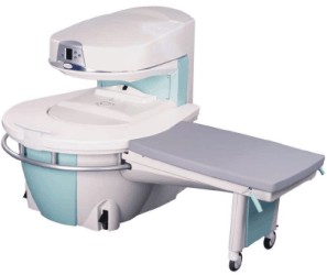
a low field open MRI scanner with permanent magnet for orthopedic use. The outstanding feature of this MRI system is a patient friendly design with 24 cm diameter, which allows the imaging of extremities and small body parts like shoulder MRI. The power consumption is around 1.3 kW and the needed minimum floor space is an area of 16 sq m.
At RSNA 2006 Hologic Inc. introduced a new dedicated extremity MRI scanner, the Opera. Manufactured by Esaote is the Opera a redesign of Esaote's 0.2 Tesla E-Scan XQ platform, which now enables complete imaging of all extremities, including hip and shoulder applications. 'Real-time positioning' reportedly speeds patient setup and reduces exam times.
Esaote North America and Hologic Inc are the U.S. distributors of this MRI device.
Device Information and Specification CLINICAL APPLICATION Dedicated extremity
SE, GE, IR, STIR, FSE, 3D CE, GE-STIR, 3D GE, ME, TME, HSE IMAGING MODES Single, multislice, volume study, fast scan, multi slab2D: 2 mm - 10 mm;
3D: 0.6 mm - 10 mm 4096 gray lvls, 256 lvls in 3D POWER REQUIREMENTS 2,0 kW; 110/220 V single phase | |  | | | |  Further Reading: Further Reading: | News & More:
|
|
| |
|  |  | Searchterm 'MRI' was also found in the following services: | | | | |
|  |  |
| |
|

The range of diagnostics and imaging systems of Siemens Medical Systems covers ultrasound, nuclear medicine, angiography, magnetic resonance, computer tomography and patient monitoring. Siemens is one of the three leading MRI manufacturers, which together account for approximately 80 percent of the MRI machines installed worldwide. Siemens currently offers the Allegra 3T MRI, which is for head scanning only, but the company will also be launching the Trio MRI, a 3T whole body scanner.
Siemens has formed partnerships with more than ten research institutions and private practitioners to define a comprehensive MRI examination and compare MR to currently established cardiovascular modalities, thereby defining optimal diagnosis and treatment.
MRI Scanners:
0.2T to 1.0T:
1.5T:
3.0T to 7.0T:
Hybrid Scanners:
Mobile Solutions:
•
MAGNETOM Espree 1.5T, MAGNETOM Avanto 1.5T and MAGNETOM ESSENZA 1.5T are also offered by Siemens on certified trailers.
Contact Information MAIL
Siemens Medical Solutions
Health Services Corporation
51 Valley Stream Parkway
Malvern, PA 19355
USA | |  | |
• View the DATABASE results for 'Siemens Medical Systems' (14).
| | |
• View the NEWS results for 'Siemens Medical Systems' (3).
| | | | |  Further Reading: Further Reading: | | Basics:
|
|
News & More:
| |
| |
|  | |  |  |  |
| |
|

Aurora Imaging Technology, Inc., a private company, is located in North Andover, Massachusetts. As a leading innovator in the breast imaging industry, the company provides with its Aurora System the detection, diagnosis, biopsy and treatment of breast cancer. Aurora Imaging Technology Inc. develops and manufactures the Aurora® 1.5T Dedicated Breast MR System, the first and only FDA approved MRI system designed specifically for breast imaging.
MRI Scanners:
Contact Information
MAIL
Aurora Imaging Technology Inc
39 High Street
North Andover, MA 01845
USA
| |  | |
• View the DATABASE results for 'Aurora Imaging Technology, Inc.' (2).
| | |
• View the NEWS results for 'Aurora Imaging Technology, Inc.' (5).
| | | | |  Further Reading: Further Reading: | News & More:
|
|
| |
|  |  | Searchterm 'MRI' was also found in the following services: | | | | |
|  |  |
| |
|
MRI guided biopsies are usually performed for lesions that are found on for example liver or breast MRI procedures and that are not seen on computed tomography, ultrasonography or mammography. The identification of cancer on breast MRI is dependent on uptake of intravenous contrast agents.
First an MRI scan, using a dedicated breast coil and biopsy guidance system is performed to found the lesion. After skin disinfection and local anesthesia, the biopsy procedure starts. Possible MR guided interventions include fine needle aspiration, core needle biopsy and vacuum-assisted biopsy (VABB) to sample tissue from the lesion; or wire localization prior to surgery for lesions that are not palpable.
See also Breast MRI.
| |  | |
• View the DATABASE results for 'Biopsy' (10).
| | |
• View the NEWS results for 'Biopsy' (6).
| | | | |  Further Reading: Further Reading: | | Basics:
|
|
News & More:
| |
| |
|  |  | Searchterm 'MRI' was also found in the following services: | | | | |
|  |  |
| |
|
During the MRI scan an augmentation of T waves is observed at fields used in standard imaging but this possible MRI side effect is completely reversible upon removal from the magnet. A field strength dependent increase in the amplitude of the ECG in rats has been observed during exposure to high homogeneous stationary magnetic fields, but this side effect is not transferable to standard imaging situations for humans.

The minimum level at which augmentation can be observed is 0.3 T and increases by higher field strength.
An augmentation in T-wave amplitude can occur instantaneously and is immediately reversible after exposure to the magnetic field ceased. There should be no abnormalities in the ECG in the later follow-up. Augmentation of the signal amplitude in the T-wave segment may result from superimposed electrical potential.
No circulatory alterations coincide with the ECG changes. Therefore, no biological risks are believed to be associated with them.
For more MRI safety information see also Contraindications
and MRI Risks. | |  | |
• View the DATABASE results for 'Cardiac Risks' (2).
| | | | |  Further Reading: Further Reading: | Basics:
|
|
| |
|  | |  |  |
|  | | |
|
| |
 | Look
Ups |
| |