 | Info
Sheets |
| | | | | | | | | | | | | | | | | | | | | | | | |
 | Out-
side |
| | | | |
|
| | | | |
Result : Searchterm 'Image Quality' found in 1 term [ ] and 44 definitions [ ] and 44 definitions [ ] ]
| | previous 36 - 40 (of 45) nextResult Pages : [1] [2 3 4 5 6 7 8 9] |  | |  | Searchterm 'Image Quality' was also found in the following services: | | | | |
| |  |
| |
|
In parallel MR imaging, a reduced data set in the phase encoding direction(s) of k-space is acquired to shorten acquisition time, combining the signal of several coil arrays. The spatial information related to the phased array coil elements is utilized for reducing the amount of conventional Fourier encoding.
First, low-resolution, fully Fourier-encoded reference images are required for sensitivity assessment. Parallel imaging reconstruction in the Cartesian case is efficiently performed by creating one aliased image for each array element using discrete Fourier transformation. The next step then is to create an full FOV image from the set of intermediate images.
Parallel reconstruction techniques can be used to improve the image quality with increased signal to noise ratio, spatial resolution, reduced artifacts, and the temporal resolution in dynamic MRI scans.
Parallel imaging algorithms can be divided into 2 main groups:
Image reconstruction produced by each coil ( reconstruction in the image domain, after Fourier transform): SENSE ( Sensitivity Encoding), PILS (Partially Parallel Imaging with Localized Sensitivity),
ASSET.
Reconstruction of the Fourier plane of images from the frequency signals of each coil ( reconstruction in the frequency domain, before Fourier transform): GRAPPA. Additional techniques include SMASH, SPEEDER™,
IPAT (Integrated Parallel Acquisition Techniques - derived of GRAPPA a k-space based technique) and mSENSE (an image based enhanced version of SENSE).
| | | | | | | | | | | | |  Further Reading: Further Reading: | | Basics:
|
|
News & More:
| |
| |
| | | Searchterm 'Image Quality' was also found in the following services: | | | | |
| | |
| |
|
| | | | | | | Further Reading: | Basics:
|
|
News & More:
| |
| |
| | | | | |
| |
|
From ISOL Technology
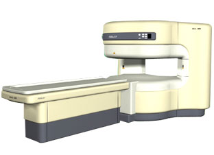
'RELAX is open type MRI system created by making up for the weakness of existing conventional MR systems and applying the strength and the application of the middle to high field MR without uncompromising the image quality.
RELAX offers you a premium mix of form, performance and functionality that are patient and user
friendly beyond comparison.
- New breed of MRI pursuing
- patients comfort'
Device Information and Specification CLINICAL APPLICATION Whole body lower than 2.4 m from the iso-center | | | |
• View the DATABASE results for 'RELAX 0.35T™' (2).
| | | | |
| | | Searchterm 'Image Quality' was also found in the following services: | | | | |
| | |
| |
|
A coil is a large inductor with a considerable dimension and a defined wavelength, commonly used in configurations for MR imaging. The frequency of the radio frequency coil is defined by the Larmor relationship. The MRI image quality depends on the signal to noise ratio (SNR) of the acquired signal from the patient. Several MR imaging coils are necessary to handle the diversity of applications. Large coils have a large measurement field, but low signal intensity and vice versa (see also coil diameter). The closer the coil to the object, the stronger the signal - the smaller the volume, the higher the SNR. SNR is very important in obtaining clear images of the human body. The shape of the coil depends on the image sampling. The best available homogeneity can be reached by choice of the appropriate coil type and correct coil positioning. Orientation is critical to the sensitivity of the RF coil and therefore the coil should be perpendicular to the static magnetic field.
RF coils can be differentiated by there function into three general categories:
The RF signal is in the range of 10 to 100 MHz. During a typical set of clinical image measurements, the entire frequency spectrum of interest is of the order 10 kHz, which is an extremely narrow band, considering that the center frequency is about 100 MHz. This allows the use of single-frequency matching techniques for coils because their inherent bandwidth always exceeds the image bandwidth. The multi turn solenoid, bird cage coil, single turn solenoid, and saddle coil are typically operated as the transmitter and receiver of RF energy. The surface and phased array coils are typically operated as a receive only coil.
See also the related poll result: ' 3rd party coils are better than the original manufacturer coils' | | | | | |
• View the DATABASE results for 'Radio Frequency Coil' (9).
| | | | | | Further Reading: | Basics:
|
|
News & More:
| |
| |
| | | Searchterm 'Image Quality' was also found in the following services: | | | | |
| | |
| |
|
SPEEDER™ is Toshiba's patented parallel imaging technology for high-speed imaging. This technique reduces examination time for MRI studies by a factor of up to three while enabling clinicians to capture high-quality images with detailed diagnostic information. With SPEEDER™, cardiac and MRA examinations are more patient-friendly and tolerable with shorter scan times and improved image quality.
See also Sensitivity Encoding. | | | |
• View the DATABASE results for 'SPEEDER™' (2).
| | | | |
| | | | |
| | | |
|
| |
 | Look
Ups |
| |


