 | Info
Sheets |
| | | | | | | | | | | | | | | | | | | | | | | | |
 | Out-
side |
| | | | |
|
| | | | |
Result : Searchterm 'Image Quality' found in 1 term [ ] and 44 definitions [ ] and 44 definitions [ ] ]
| | previous 16 - 20 (of 45) nextResult Pages : [1] [2 3 4 5 6 7 8 9] |  | |  | Searchterm 'Image Quality' was also found in the following services: | | | | |
| |  |
| |
|
(FOV) Defined as the size of the two or three dimensional spatial encoding area of the image. Usually defined in units of mm². The FOV is the square image area that contains the object of interest to be measured. The smaller the FOV, the higher the resolution and the smaller the voxel size but the lower the measured signal.
Useful for decreasing the scantime is a field of view different in the frequency and phase encoding directions ( rectangular field of view - RFOV).
The magnetic field homogeneity decreases as more tissue is imaged (greater FOV). As a result the precessional frequencies change across the imaging volume. That can be a problem for fat suppression imaging. This fat is precessing at the expected frequency only in the center of the imaging volume. E.g. frequency specific fat saturation pulses become less effective when the field of view is increased. It is best to use smaller field of views when applying fat saturation pulses.

Image Guidance
Smaller FOV required higher gradient strength and concludes low signal. Therefore you have to find a compromise between these factors.
The right choice of the field of view is important for MR image quality. When utilizing small field of views and scanning at a distance from the isocenter (more problems with artifacts) it is obviously important to ensure that the region of interest is within the scanning volume.
A smaller FOV in one direction is available with the function rectangular field of view (RFOV).
See also Field Inhomogeneity Artifact. | | | | | | | | | | | | |  Further Reading: Further Reading: | | Basics:
|
|
News & More:
| |
| |
| | | Searchterm 'Image Quality' was also found in the following services: | | | | |
| | |
| |
|
A measure of the geometrical relationship of the RF coil and the object being studied. It affects the efficiency of exciting the object and detecting MR signals, thereby affecting the signal to noise ratio and image quality. Achieving a high filling factor requires fitting the coil closely to the object, thus potentially decreasing patient comfort. | | | | | | | Further Reading: | News & More:
|
|
| |
| | | | | |
| |
|
The principal advantage of MRI at high field is the increase in signal to noise ratio. This can be used to improve anatomic and/or temporal resolution and reduce scan time while preserving image quality. MRI devices for whole body imaging for human use are available up to 3 tesla (3T). Functional MRI ( fMRI) and MR spectroscopy ( MRS) benefit significantly. In addition, 3T machines have a great utility in applications such as TOF MRA and DTI. Higher field strengths are used for imaging of small parts of the body or scientific animal experiments. Higher contrast may permit reduction of gadolinium doses and, in some cases, earlier detection of disease.
Using high field MRI//MRS, the RF-wavelength and the dimension of the human body complicating the development of MR coils. The absorption of RF power causes heating of the tissue. The energy deposited in the patient's tissues is fourfold higher at 3T than at 1.5T. The specific absorption rate (SAR) induced temperature changes of the human body are the most important safety issue of high field MRI//MRS.
Susceptibility and chemical shift dispersion increase like T1, therefore high field MRI occasionally exhibits imaging artifacts. Most are obvious and easily recognized but some are subtle and mimic diseases. A thorough understanding of these artifacts is important to avoid potential pitfalls. Some imaging techniques or procedures can be utilized to remove or identify artifacts. See also Diffusion Tensor Imaging.
See also the related poll result: ' In 2010 your scanner will probably work with a field strength of' | | | | | |
• View the DATABASE results for 'High Field MRI' (16).
| | |
• View the NEWS results for 'High Field MRI' (9).
| | | | | | Further Reading: | Basics:
|
|
News & More:
| |
| |
| | | Searchterm 'Image Quality' was also found in the following services: | | | | |
| | |
| |
|
The image resolution is the level of detail of an image and a measurement of image quality. Higher resolution means more image detail, for example when two structures 1 mm apart are distinguishable in an image, this picture has a higher resolution than an image where they are not to distinguish.
More data points in an MR image (with same FOV) will decrease the pixel size, but not accurately improve the resolution because the different MRI sequences influence the contrast and the discernment of different tissues.
With high contrast and optimal signal to noise ratio, the image resolution is depend on FOV and number of data points of a picture, but T2* effects have an additional influence. | | | |
• View the DATABASE results for 'Image Resolution' (9).
| | |
• View the NEWS results for 'Image Resolution' (1).
| | | | | | Further Reading: | Basics:
|
|
News & More:
| |
| |
| | | Searchterm 'Image Quality' was also found in the following services: | | | | |
| | |
| |
|
From Philips Medical Systems;
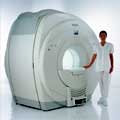
Philips Infinion 1.5 T is designed to maximize the efficiency and quality of patient care. Developed with the patient in mind, the Infinion is the shortest and most open 1.5T scanner available. The unique 'ultra short' 1.4 m magnet assures patient comfort and acceptance without compromising image quality and clinical performance.
Device Information and Specification
CLINICAL APPLICATION
Whole body
CONFIGURATION
Ultra short bore
Head, head / neck, integrated C-spine, L/T spine array, small large GP coils, body flex array, torso pelvis array, breast array, endocavitary, shoulder array, lower extremity, hand / wrist, cardiac, PV array
SE, TSE, SS TSE, EPI, IR, STIR, FLAIR, FFE, TFE, T1 TFE, T2 TFE, Presat, Fatsat, MTC, Diff-opt., Angiography: PCA, MCA, TOF
IMAGING MODES
Single slice, single volume, multi slice, multi volume
80 images/sec std.; up to320 opt.@256
H*W*D
233 (lead fitted) x 198 x 140 cm
POWER REQUIREMENTS
400/480 V
COOLING SYSTEM TYPE
Closed loop, chilled water
| | | |
• View the DATABASE results for 'Infinion 1.5T™' (2).
| | | | |
| | | | |
| | | |
|
| |
 | Look
Ups |
| |


