 | Info
Sheets |
| | | | | | | | | | | | | | | | | | | | | | | | |
 | Out-
side |
| | | | |
|
| | | | |
Result : Searchterm 'Hardware' found in 1 term [ ] and 20 definitions [ ] and 20 definitions [ ] ]
| | previous 16 - 20 (of 21) nextResult Pages : [1] [2 3 4 5] |  | |  | Searchterm 'Hardware' was also found in the following services: | | | | |
| |  |
| |
|
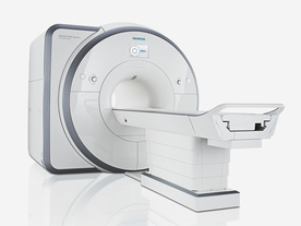
From Siemens Medical Systems;
Received FDA clearance in 2012.
The MAGNETOM Spectra is a cost-optimized high field MRI system with Tim 4G and Dot technologies. The system consumes less energy compared to other 3 Tesla scanners. The magnet-cooling helium is contained in a closed loop, which prevents the gas from escaping and reduces the need for refills. TimTX includes innovative techniques in the radio frequency excitation hardware as well as new application and processing features enabling uniform RF distribution in all body regions.
Device Information and Specification
CLINICAL APPLICATION
Whole Body
Head, spine, torso/ body coil, neurovascular, neck and multi-purpose flex coils. Peripheral vascular, breast, shoulder, knee, wrist, foot//ankle, endorectal optional.
Chemical shift imaging, single voxel spectroscopy
DIMENSION H*W*D (gantry included)
173 x 231 x 219 cm
COOLING SYSTEM
Water; single cryogen, 2 stage refrigeration
Passive, active; first order standard, second order optional
POWER REQUIREMENTS
380 / 400 / 420 / 440 / 460 / 480 V, 3-phase + ground; connection value with chiller 100 kvA /without chiller 60 kVA
| | | | | |
| | | Searchterm 'Hardware' was also found in the following services: | | | | |
| | |
| |
|

It is important to remember when working around a superconducting magnet that the magnetic field is always on. Under usual working conditions the field is never turned off. Attention must be paid to keep all ferromagnetic items at an adequate distance from the magnet. Ferromagnetic objects which came accidentally under the influence of these strong magnets can injure or kill individuals in or nearby the magnet, or can seriously damage every hardware, the magnet itself, the cooling system, etc..
See MRI resources Accidents.
The doors leading to a magnet room should be closed at all times except when entering or exiting the room. Every person working in or entering the magnet room or adjacent rooms with a magnetic field has to be instructed about the dangers. This should include the patient, intensive-care staff, and maintenance-, service- and cleaning personnel, etc..
The 5 Gauss limit defines the 'safe' level of static magnetic field exposure. The value of the absorbed dose is fixed by the authorities to avoid heating of the patient's tissue and is defined by the specific absorption rate.
Leads or wires that are used in the magnet bore during imaging procedures, should not form large-radius wire loops. Leg-to-leg and leg-to-arm skin contact should be prevented in order to avoid the risk of burning due to the generation of high current loops if the legs or arms are allowed to touch. The patient's skin should not be in contact with the inner bore of the magnet.
The outflow from cryogens like liquid helium is improbable during normal operation and not a real danger for patients.
The safety of MRI contrast agents is tested in drug trials and they have a high compatibility with very few side effects. The variations of the side effects and possible contraindications are similar to X-ray contrast medium, but very rare. In general, an adverse reaction increases with the quantity of the MRI contrast medium and also with the osmolarity of the compound.
See also 5 Gauss Fringe Field, 5 Gauss Line, Cardiac Risks, Cardiac Stent, dB/dt, Legal Requirements, Low Field MRI, Magnetohydrodynamic Effect, MR Compatibility, MR Guided Interventions, Claustrophobia, MRI Risks and Shielding. | | | | | | | | |
• View the DATABASE results for 'MRI Safety' (42).
| | |
• View the NEWS results for 'MRI Safety' (13).
| | | | |  Further Reading: Further Reading: | | Basics:
|
|
News & More:
| |
| |
| | | | | |
| |
|
Quick Overview Please note that there are different common names for this artifact.
DESCRIPTION
Striped ghosts with a shift of half the field of view
Machine imperfection-based artifacts manifest themselves due to the fact that the odd k-space lines are acquired in a different direction than the even k-space lines. Slight differences in timing result in shifts of the echo in the acquisition window. By the shift theorem, such shifts in the time domain data then produce linear phase differences in the frequency domain data.
Without correction, such phase differences in every second line produce striped ghosts with a shift of half the field of view, so-called Nyquist ghosts. Shifts in the applied magnetic field can also produce similar (but constant in amplitude) ghosts.
This artifact is commonly seen in an EPI image and can arise from both, hardware and sample imperfections.
A further source of machine-based artifact arises from the need to acquire the signal as quickly as possible. For this reason the EPI signal is often acquired during times when the gradients are being switched. Such sampling effectively means that the k-space sampling is not uniform, resulting in ringing artifacts in the image.

Image Guidance
Such artifacts can be minimized by careful setup of the spectrometer and/or correction of the data. For this reasons reference data are often collected, either as a separate scan or embedded in the imaging data.
The non-uniform sampling can be removed by knowing the form of the gradient switching. It is possible to regrid the data onto a uniform k-space grid. | | | |
• View the DATABASE results for 'Machine Imperfection Artifact' (2).
| | | | | | Further Reading: | Basics:
|
|
| |
| | | Searchterm 'Hardware' was also found in the following services: | | | | |
| | |
| |
|
Quick Overview
NAME
Metal, susceptibility
Ferromagnetic metal will cause a magnetic field inhomogeneity, which in turn causes a local signal void, often accompanied by an area of high signal intensity, as well as a distortion of the image.
They create their own magnetic field and dramatically alter precession frequencies of protons in the adjacent tissues. Tissues adjacent to ferromagnetic components become influenced by the induced magnetic field of the metal hardware rather than the parent field and, therefore, either fail to precess or do so at a different frequency and hence do not generate useful signal. Two components contribute to susceptibility artifact, induced magnetism in the ferromagnetic component itself and induced magnetism in protons adjacent to the component. Artifacts from metal may have varied appearances on MRI scans due to different type of metal or configuration of the piece of metal.
The biocompatibility of metallic alloys, stainless steel, cobalt chrome and titanium alloy is based on the presence of a constituent element within the alloy that has the ability to form an adherent oxide coating that is stable, chemically inert and hence biocompatible. In relation to imaging titanium alloys are less ferromagnetic than both cobalt and stainless steel, induce less susceptibility artifact and result in less marked image degradation.
Image Guidance
| | | |
• View the DATABASE results for 'Metal Artifact' (2).
| | | | | | Further Reading: | | Basics:
|
|
News & More:
| |
| |
| | | Searchterm 'Hardware' was also found in the following services: | | | | |
| | |
| |
|
A pulse sequence is a preselected set of defined RF and gradient pulses, usually repeated many times during a scan, wherein the time interval between pulses and the amplitude and shape of the gradient waveforms will control NMR signal reception and affect the characteristics of the MR images. Pulse sequences are computer programs that control all hardware aspects of the MRI measurement process.
Usual to describe pulse sequences, is to list the repetition time (TR), the echo time (TE), if using inversion recovery, the inversion time (TI) with all times given in milliseconds, and in case of a gradient echo sequence, the flip angle. For example, 3000/30/1000 would indicate an inversion recovery pulse sequence with TR of 3000 msec., TE of 30 msec., and TI of 1000 msec.
Specific pulse sequence weightings are dependent on the field strength, the manufacturer and the pathology.
See also Interpulse Times. | | | |
• View the DATABASE results for 'Pulse Sequence' (96).
| | |
• View the NEWS results for 'Pulse Sequence' (1).
| | | | | | Further Reading: | Basics:
|
|
News & More:
| |
| |
| | | | |
| | | |
|
| |
 | Look
Ups |
| |


