Magnetic Resonance - Technology Information Portal Welcome to MRI Technology
Info
  Sheets

Out-
      side


 'Gating' 
SEARCH FOR    
 
  2 3 5 A B C D E F G H I J K L M N O P Q R S T U V W X Y Z
Result : Searchterm 'Gating' found in 4 terms [] and 63 definitions []
previous     16 - 20 (of 67)     next
Result Pages : [1]  [2 3 4 5 6 7 8 9 10 11 12 13 14]
Searchterm 'Gating' was also found in the following services: 
spacer
News  (4)  Resources  (2)  Forum  (1)  
 
Virgo™InfoSheet: - Devices -
Intro, 
Types of Magnets, 
Overview, 
etc.MRI Resource Directory:
 - Devices -
 
www.canamglobal.com/mri/virgomri.html From Millennium Technology Inc. 
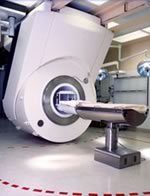
This open C-shaped MRI system eases patient comfort and technologist maneuverability. This low cost scanner is build for a wide range of applications. The Virgo™ patient table is detachable and moves on easy rolling castors. Able to accommodate patient weights up to 160 kg, the tabletop has a range of motion of 30 cm in the lateral direction and 90cm in the longitudinal direction. Images generated with this scanner can only be viewed (without data loss) on Millennium's proprietary viewing software.
Device Information and Specification
CLINICAL APPLICATION
Whole body
CONFIGURATION
C shaped
SYNCHRONIZATION
Standard cardiac gating, ECG/peripheral, respiratory gating
PULSE SEQUENCES
2D Spin Echo (single and multi-echo), 2D Inversion Recovery, 2D Sequential and 3D Volume Gradient Echo, 2D and 3D Spoiled Gradient Echo
IMAGING MODES
Localizer, single slice, multislice, volume, fast, POMP, multi slab, cine, slice and frequency zip, extended dynamic range, tailored RF
TR
steps of 1 msec
TE
steps of 1 msec
SINGLE/MULTI SLICE
Simultaneous scan and reconstruction;; 100 images/second reconstruction
400 mm
2D : 2 mm; 3D : 0.5 mm
MEASURING MATRIX
512x512
PIXEL INTENSITY
256 gray levels
MAGNET TYPE
Permanent
BORE DIAMETER
or Vertical Gap
44 cm
STRENGTH
15 mT/m
5-GAUSS FRINGE FIELD
3 m/3 m
Passive
spacer
Searchterm 'Gating' was also found in the following services: 
spacer
Radiology  (5) Open this link in a new windowUltrasound  (7) Open this link in a new window
iMotion™ 1.5 Tesla MagnetInfoSheet: - Devices -
Intro, 
Types of Magnets, 
Overview, 
etc.MRI Resource Directory:
 - Devices -
 
www.imris.com/ The mobile intraoperative iMotion system produces real-time images used for MR guided surgery and offers functional magnetic resonance imaging, MR spectroscopy, perfusion imaging, and diffusion weighted imaging capabilities.
The iMotion 1.5 T magnet moves to the patient, gliding in and out of place as needed, without affecting surgical, anesthetic, and nursing management.

See also Intraoperative Magnetic Resonance Imaging, MR Guided Interventions.
Device Information and Specification
CLINICAL APPLICATION
Whole body
CONFIGURATION
Mobile compact
Whole body, intra-operative head, neck volume, atlas head//neck vascular quadrature phased array, spine quadrature, C/T/L spine phased array, small joint, large joint, TMJ bilateral, shoulder phased array, extremity quadrature volume, wrist, hand quadrature, general purpose flexible, pelvis/abdomen phased array, body quadrature, phased array flexible, breast bilateral
SYNCHRONIZATION
Standard cardiac gating, ECG/peripheral, respiratory gating
PULSE SEQUENCES
SE, IR, TSE, EPI, Diffusion, Perfusion, Angiography
IMAGING MODES
Localizer, single slice, multislice, volume
Active
spacer

• View the DATABASE results for 'iMotion™ 1.5 Tesla Magnet' (2).Open this link in a new window

MRI Resources 
Bioinformatics - PACS - Service and Support - Jobs pool - Pathology - Lung Imaging
 
Arrhythmia Rejection
 
With this method irregular RR intervals in cardiac gating during cardiovascular imaging are rejected and then repeated to improve the image quality, whereby the cardiac frequency is used as a basis of the normal heart rate.
The RR interval window determines the percentage variation of the heart rate. Variations of the acquired data outside the window are rejected and not used in the image reconstruction. Also one interval after the arrhytmic beat will be rejected.
Arrhythmia rejection may be inappropriate for patients with certain pathologies, because if the RR interval is constant long, short, long, - all intervals would be rejected. Also a disadvantage is the time consume, but in some cases this function is mandatory, e.g. for diverse retrospective triggered sequences.
spacer
 
Further Reading:
  Basics:
A Guide To Cardiac Imaging
   by www.simplyphysics.com    
  News & More:
Irregular heartbeat may lead to silent strokes
Wednesday, 5 November 2014   by www.techtimes.com    
Searchterm 'Gating' was also found in the following services: 
spacer
News  (4)  Resources  (2)  Forum  (1)  
 
ArtifactForum -
related threadsInfoSheet: - Artifacts - 
Case Studies, 
Reduction Index, 
etc.MRI Resource Directory:
 - Artifacts -
 
An image artifact is a structure not normally present but visible as a result of a limitation or malfunction in the hardware or software of the MRI device, or in other cases a consequence of environmental influences as heat or humidity or it can be caused by the human body (blood flow, implants etc.). The knowledge of MRI artifacts (brit. artefacts) and noise producing factors is important for continuing maintenance of high image quality. Artifacts may be very noticeable or just a few pixels out of balance but can give confusing artifactual appearances with pathology that may be misdiagnosed.
Changes in patient position, different pulse sequences, metallic artifacts, or other imaging variables can cause image distortions, which can be reduced by the operator; artifacts due to the MR system may require a service engineer.
Many types of artifacts may occur in magnetic resonance imaging. Artifacts in magnetic resonance imaging are typically classified as to their basic principles, e.g.:
•
Physiologic (motion, flow)
•
Hardware (electromagnetic spikes, ringing)
•
Inherent physics (chemical shift, susceptibility, metal)

Several techniques are developed to reduce these artifacts (e.g. respiratory compensation, cardiac gating, eddy current compensation) but sometimes these effects can also be exploited, e.g. for flow measurements.

See also the related poll result: 'Most outages of your scanning system are caused by failure of'
spacer

• View the DATABASE results for 'Artifact' (166).Open this link in a new window

 
Further Reading:
  Basics:
ARTEFACT VERSUS ARTIFACT
Saturday, 26 January 2002   by www.worldwidewords.org    
  News & More:
MRI results affected by movement? MIT researchers have an AI-powered solution
Friday, 25 August 2023   by healthimaging.com    
Magnetic eyelashes: A new source of MRI artifacts
Wednesday, 24 July 2019   by medicalxpress.com    
On the Horizon - Next Generation MRI
Wednesday, 23 October 2013   by thefutureofthings.com    
Searchterm 'Gating' was also found in the following services: 
spacer
Radiology  (5) Open this link in a new windowUltrasound  (7) Open this link in a new window
Blood Pool AgentsInfoSheet: - Contrast Agents - 
Intro, Overview, 
Characteristics, 
Types of, 
etc.MRI Resource Directory:
 - Contrast Agents -
 
Blood pool agents (intravascular contrast agents) remain in the blood for a prolonged time compared with conventional contrast agents, which diffuse quickly into the interstitial space. Magnetic resonance angiography (MRA), cardiovascular imaging, or contrast enhanced MRIs are possible over an hour or more. This advantage over conventional MRI contrast media allows also higher resolution MRA of several territories using respiratory or cardiac gating techniques with a single contrast bolus.
Different types of blood pool contrast agents:
Blood pool MRI contrast agents with their longer intravascular circulation can be designed to be targeted to necrotic myocardium, to assess myocardial viability, or tumor directed to provide better diagnostic information for various tumors. A disadvantage of the use of blood pool agents for MRA is that the separation of arteries and veins is more difficult because they are present in both and the overlapping of those vessels is disturbing. This can be solved by e.g. different MIP segmentation algorithms.

See also Necrosis Avid Contrast Agent, Tumor Specific Agents, Feruglose, Gadofosveset Trisodium (Vasovist), Ultrasmall Superparamagnetic Iron Oxide and Contrast Medium.
spacer

• View the DATABASE results for 'Blood Pool Agents' (16).Open this link in a new window


• View the NEWS results for 'Blood Pool Agents' (1).Open this link in a new window.
 
Further Reading:
  Basics:
Ablavar Prescribing Information
   by www.ablavar.com    
Lantheus Medical Imaging, Inc. Launches ABLAVAR™ (Gadofosveset Trisodium), a New Diagnostic Magnetic Resonance Angiography Agent
Wednesday, 20 January 2010   by www.radiopharm.com    
Blood-Pool Imaging Using Technetium-99m-Labeled Liposomes(.pdf)
   by jnm.snmjournals.org    
  News & More:
Multimodal Nanoparticles for Quantitative Imaging(.pdf)
Tuesday, 13 December 2011   by alexandria.tue.nl    
MAGNETIC RESONANCE IMAGING OF FOCAL LIVER LESIONS(.pdf)
2002
MRI Resources 
Shielding - Pathology - Jobs pool - General - Universities - Corporations
 
previous      16 - 20 (of 67)     next
Result Pages : [1]  [2 3 4 5 6 7 8 9 10 11 12 13 14]
 Random Page
 
Share This Page
FacebookTwitterLinkedIn

MR-TIP    
Community   
User
Pass
Forgot your UserID/Password ?    


Next big thing in MRI will be :
AI 
remote operator 
personalized protocols 
helium-free 
molecular MRI 
portable MRI 

Look
      Ups


MR-TIP.com uses cookies! By browsing MR-TIP.com, you agree to our use of cookies.

Magnetic Resonance - Technology Information Portal
Member of SoftWays' Medical Imaging Group - MR-TIP • Radiology-TIP • Medical-Ultrasound-Imaging • 
Copyright © 2003 - 2024 SoftWays. All rights reserved. [ 25 April 2024]
Terms of Use | Privacy Policy | Advertising
 [last update: 2024-02-26 03:41:00]